A detailed illustration of a chick embryo in an open humidity chamber, viewed under a microscope, showcasing its vascular system and developing organs.

Unlocking Life's Secrets: A Beginner's Guide to Chick Embryo Development

"From Tiny Egg to Thriving Chick: Discovering the Wonders of Embryo Development with an Innovative Ex-Ovo Technique"


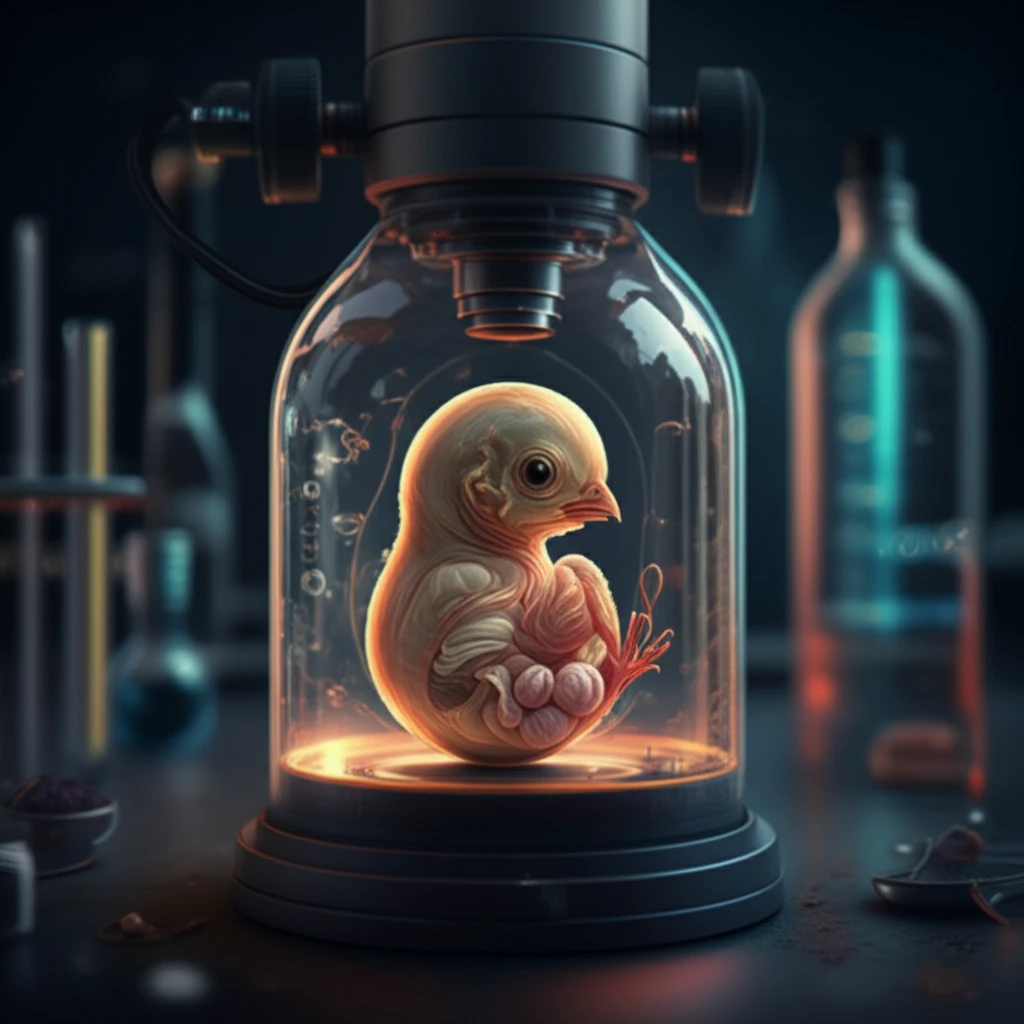
The study of life's beginnings – the development of an embryo – has long been a captivating area of scientific inquiry. For generations, researchers have sought ways to unlock the secrets hidden within the earliest stages of life. One of the most fascinating models for this exploration has been the humble chicken embryo. Enclosed within an egg, these embryos offer a unique opportunity to study development, but also present some challenges. Traditional methods often involve complex procedures, but there's a simpler, more accessible way to witness this incredible transformation.

This article introduces an optimized 'ex ovo' culturing technique, a method that allows you to observe chick embryos as they grow, right before your eyes. Forget complicated setups; this technique simplifies the process, making it accessible for everyone, from budding biology enthusiasts to seasoned researchers. We'll delve into the ease of this method, highlighting how it provides a clear view of embryonic development, from the initial stages to the formation of vital organs.

Join us as we journey into the world of chick embryos, discovering how you too can unlock the secrets of life's most fundamental processes. This isn't just about science; it's about witnessing the marvels of nature and understanding the incredible journey from a single cell to a fully formed creature. Get ready to be amazed by the wonders of embryo development!

The Ex Ovo Advantage: Witnessing Life Unfold

A detailed illustration of a chick embryo in an open humidity chamber, viewed under a microscope, showcasing its vascular system and developing organs.

Traditional methods of studying chick embryos often involve either windowing the eggshell or using complex ex ovo setups. Windowing allows a peek inside but can be limiting, particularly in the later stages when the embryo grows significantly. Complex ex ovo methods, while offering more control, can be technically challenging and often require specialized equipment. Our optimized ex ovo technique offers a solution, providing a clear and accessible way to observe chick embryo development.

The core of this technique revolves around a simple, yet ingenious approach: creating a controlled environment for the embryo outside the egg. This allows for easy observation under a microscope and facilitates manipulations without the constraints of the eggshell. Here’s how the ex ovo method opens the door to deeper understanding:

  • Unobstructed View: Unlike windowing, this method provides a complete view of the developing embryo at all stages.
  • Ease of Use: The setup is straightforward, requiring no specialized equipment, making it ideal for both educational and research settings.
  • Manipulation Friendly: It provides easy access for manipulations such as introducing drugs and dyes.
  • Developmental Insights: Allows observation of the later stages of development, including ossification and feather growth.
With the ex ovo method, you can witness the incredible transformation from a few cells into a complex organism. The complete embryo and its extraembryonic membranes and blood vessels are readily visible, providing a wealth of information on developmental biology.

Embark on Your Embryo Adventure!

The journey through embryo development is a captivating exploration of life's most fundamental processes. With the optimized ex ovo technique, the mysteries of chick embryo development are within easy reach. Whether you're a student eager to learn or a researcher seeking innovative methods, this approach offers a clear, accessible path to discovery. So, gather your materials, follow the steps, and prepare to witness the extraordinary unfolding of life!

About this Article -

This article was crafted using a human-AI hybrid and collaborative approach. AI assisted our team with initial drafting, research insights, identifying key questions, and image generation. Our human editors guided topic selection, defined the angle, structured the content, ensured factual accuracy and relevance, refined the tone, and conducted thorough editing to deliver helpful, high-quality information.See our About page for more information.

Everything You Need To Know

1

What is the 'ex ovo' culturing technique, and why is it considered an advancement in studying chick embryo development?

The 'ex ovo' culturing technique is an optimized method for observing chick embryo development outside of the egg. It's an advancement because it offers a clear, unobstructed view of the developing embryo at all stages, unlike traditional methods like windowing, which can be limiting. Furthermore, it simplifies the observation process and manipulations, making it accessible for a wider range of users without needing specialized equipment that may be required by other complex 'ex ovo' setups. This enables easier observation of the later stages of development, including ossification and feather growth.

2

How does the 'ex ovo' method specifically enhance the study of chick embryo development compared to traditional methods like 'windowing'?

The 'ex ovo' method provides several advantages over traditional 'windowing' techniques. 'Windowing' involves creating a small opening in the eggshell to observe the embryo, which can be restrictive, particularly as the embryo grows. In contrast, the 'ex ovo' method allows for a complete and unobstructed view of the entire chick embryo and its extraembryonic membranes and blood vessels throughout all developmental stages. Furthermore, it facilitates manipulations, such as introducing drugs and dyes, more easily. This comprehensive view and ease of access make it ideal for detailed studies of developmental biology.

3

What are the key benefits of using the optimized 'ex ovo' technique, especially in terms of accessibility and ease of use for both educational and research purposes?

The optimized 'ex ovo' technique has several key benefits. Its simplicity and ease of setup make it highly accessible for educational settings, allowing students to directly observe chick embryo development without complex procedures. For research purposes, the technique provides an unobstructed view of the developing embryo, which is essential for detailed analysis. The ability to easily manipulate the embryo, such as introducing drugs and dyes, allows for controlled experiments. Together, these benefits make it a valuable tool for both teaching and advancing our understanding of developmental biology.

4

How does observing chick embryo development using the 'ex ovo' method contribute to our understanding of fundamental processes in developmental biology?

Observing chick embryo development through the 'ex ovo' method provides a wealth of information about fundamental processes in developmental biology. By offering a clear view of the embryo and its extraembryonic membranes and blood vessels, researchers can study organ formation, cell differentiation, and the effects of various factors on development. This insight is crucial for understanding the complex journey from a single cell to a fully formed organism. Further studies into ossification and feather growth can also be easily done. This deeper understanding can then be applied to broader biological contexts.

5

Beyond basic observation, what kind of experimental manipulations are facilitated by the 'ex ovo' technique, and how might these manipulations lead to new discoveries in developmental biology or related fields?

The 'ex ovo' technique facilitates various experimental manipulations, such as the introduction of drugs and dyes, allowing researchers to study the effects of specific substances on chick embryo development. These manipulations can lead to new discoveries in developmental biology by revealing the roles of different genes and signaling pathways in organ formation, cell differentiation, and other developmental processes. For example, researchers can use this technique to test the effects of potential teratogens or to study the mechanisms of tissue regeneration, potentially leading to advancements in medicine or biotechnology.

Newsletter Subscribe

Subscribe to get the latest articles and insights directly in your inbox.