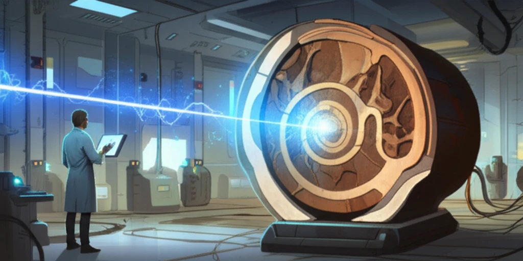
Illustration of neutron beam passing through a fossil, revealing its internal structure.

Unlocking Hidden Worlds: How Neutron Imaging is Revolutionizing Life Sciences

"Dive into the cutting-edge world of neutron imaging and discover how this revolutionary technique is transforming research in biology, palaeontology, and beyond."


In the realm of scientific discovery, innovation constantly pushes the boundaries of what's possible. Among these advancements, neutron imaging stands out as a transformative technique, particularly within the life sciences. It allows scientists to peer inside biological specimens without causing damage, revealing hidden structures and compositions that were once inaccessible.

Imagine being able to examine the internal structure of a fossil without breaking it open or study the water distribution within a plant's roots in real-time. This is the power of neutron imaging. This non-destructive approach, which offers a unique perspective, complements traditional methods like X-ray and magnetic resonance imaging, providing unparalleled insights into the intricate workings of life.

This article delves into the groundbreaking applications of neutron imaging in life sciences, from biology and palaeontology to agriculture and dentistry. It explores how this technology works, highlights its advantages, and showcases its potential to revolutionize research across various scientific fields. Get ready to discover how neutron imaging is unlocking hidden worlds, one neutron at a time.

Unveiling the Secrets Within: How Neutron Imaging Works

Illustration of neutron beam passing through a fossil, revealing its internal structure.

At its core, neutron imaging operates on a simple, yet elegant principle: sending a beam of neutrons through a sample and observing how they interact. Neutrons, as highly penetrating particles, can pass through matter with minimal disruption. This characteristic makes them ideal for examining the internal features of various materials, including biological samples.

Unlike X-rays, which interact primarily with electrons, neutrons interact with the atomic nuclei. This difference provides unique advantages. Neutrons are particularly sensitive to hydrogen, making them exceptional at visualizing water content and other hydrogen-rich substances within samples. The degree to which neutrons are absorbed or scattered provides information about the internal structure and composition of the sample.

  • Penetrating Power: Neutrons can pass through materials without causing significant structural changes.
  • Hydrogen Sensitivity: Neutrons interact strongly with hydrogen, making them perfect for studying water distribution and organic matter.
  • Non-Destructive: Samples remain intact throughout the imaging process, preserving their integrity.
  • Complementary Technique: It provides unique insights that complement other imaging methods like X-ray and MRI.
The data collected from neutron imaging is then processed to create detailed 2D or 3D images, revealing the internal structure of the sample. Scientists can analyze these images to gain insights into the composition, density, and distribution of various components within the sample, which ultimately help understand its internal features.

The Future of Discovery: The Impact of Neutron Imaging

Neutron imaging is rapidly becoming an indispensable tool in life sciences, offering unprecedented opportunities for discovery. Its ability to reveal the unseen, combined with its non-destructive nature, positions it as a key technology for researchers worldwide. As technology advances, the scope of neutron imaging applications will only expand, promising exciting advancements in our understanding of the natural world and the intricate systems that govern life. The potential of neutron imaging is vast, and the future of discovery looks brighter than ever.

About this Article -

This article was crafted using a human-AI hybrid and collaborative approach. AI assisted our team with initial drafting, research insights, identifying key questions, and image generation. Our human editors guided topic selection, defined the angle, structured the content, ensured factual accuracy and relevance, refined the tone, and conducted thorough editing to deliver helpful, high-quality information.See our About page for more information.

This article is based on research published under:

DOI-LINK: 10.1111/jmi.12761, Alternate LINK

Title: Exploring The Potential Of Neutron Imaging For Life Sciences On Imat

Subject: Histology

Journal: Journal of Microscopy

Publisher: Wiley

Authors: G. Burca, S. Nagella, T. Clark, D. Tasev, I.A. Rahman, R.J. Garwood, A.R.T. Spencer, M.J. Turner, J.F. Kelleher

Published: 2018-10-15

Everything You Need To Know

1

What makes neutron imaging stand out from other imaging techniques used in life sciences?

Neutron imaging distinguishes itself primarily through its non-destructive nature and its unique interaction with matter. Unlike X-ray imaging, which interacts with electrons, neutron imaging interacts with the atomic nuclei. This interaction provides high sensitivity to hydrogen, making it particularly effective for visualizing water content and organic matter within biological samples. This is crucial in studying biological specimens without causing damage, thereby preserving the integrity of the sample for further analysis. Other methods, like MRI, offer different insights, but neutron imaging's capacity to penetrate deeply and highlight specific elements like hydrogen provides a complementary and often more detailed view.

2

How does neutron imaging work to reveal the internal structures of biological samples?

Neutron imaging works by directing a beam of neutrons through a sample. As these neutrons pass through, they interact with the atomic nuclei within the sample. The degree to which the neutrons are absorbed or scattered provides information about the internal structure and composition of the sample. Because neutrons are highly penetrating, they can pass through the material with minimal disruption, making them ideal for examining internal features. The data collected is then processed to create detailed 2D or 3D images, allowing scientists to analyze the composition, density, and distribution of various components within the sample.

3

Why is the sensitivity of neutron imaging to hydrogen so important in biological research?

The sensitivity of neutron imaging to hydrogen is crucial because hydrogen is a major component of water and organic matter, both of which are fundamental to biological systems. This sensitivity allows scientists to visualize water distribution within a plant's roots or study the composition of organic tissues in a fossil, offering insights that would be difficult or impossible to obtain with other imaging techniques. This capability provides a deeper understanding of biological processes and structures at a molecular level.

4

What are some limitations of neutron imaging, and how do researchers address them to gain a more complete understanding?

While neutron imaging offers unique advantages, it is not without limitations. One notable limitation is access to neutron sources, which are typically large research facilities. Furthermore, the spatial resolution of neutron imaging might not always match that of other techniques like electron microscopy. To address these limitations, researchers often combine neutron imaging with other methods such as X-ray imaging, MRI, and microscopy. By integrating data from multiple sources, they can create a more comprehensive picture of the sample, leveraging the strengths of each technique while overcoming individual limitations. This multidisciplinary approach ensures a more thorough and accurate analysis.

5

What are the broad implications of neutron imaging for the future of biological and palaeontological research?

The broad implications of neutron imaging for the future of biological and palaeontological research are immense. Its non-destructive nature and unique ability to visualize internal structures and compositions will enable researchers to study samples in unprecedented detail. In biology, this could lead to breakthroughs in understanding plant physiology, disease mechanisms, and drug delivery systems. In palaeontology, it allows for the examination of fossils without damaging them, potentially revealing new insights into the evolution of life on Earth. As technology advances, the accessibility and capabilities of neutron imaging will continue to expand, promising exciting advancements in our understanding of the natural world and the intricate systems that govern life.

Newsletter Subscribe

Subscribe to get the latest articles and insights directly in your inbox.