Glowing green light in foot veins, symbolizing improved blood flow and healing through IG-FI technology.

Unlocking Healing: How New Imaging Tech Could Save Limbs

"Ischemic Foot Perfusion: A Breakthrough in Vascular Health?"


For individuals facing the threat of limb loss due to ischemia, the quest for effective treatments is paramount. Ischemic foot, a condition characterized by reduced blood flow, poses significant challenges to healing and overall quality of life. However, a promising innovation has emerged in the form of indocyanine green fluorescence imaging (IG-FI).

IG-FI is emerging as a transformative tool, providing clinicians with unprecedented insights into perfusion dynamics before and after revascularization procedures. As detailed in a recent study presented at the 33rd Annual Meeting of the French Society for Vascular and Endovascular Surgery, IG-FI offers the potential to revolutionize the assessment and management of ischemic feet.

This article delves into the science behind IG-FI, exploring its applications, benefits, and potential impact on patient outcomes. We will simplify the key findings of the study, making the complex research accessible to a broader audience, including those directly affected by ischemic conditions and their loved ones.

The Science of IG-FI: Seeing the Unseen

Glowing green light in foot veins, symbolizing improved blood flow and healing through IG-FI technology.

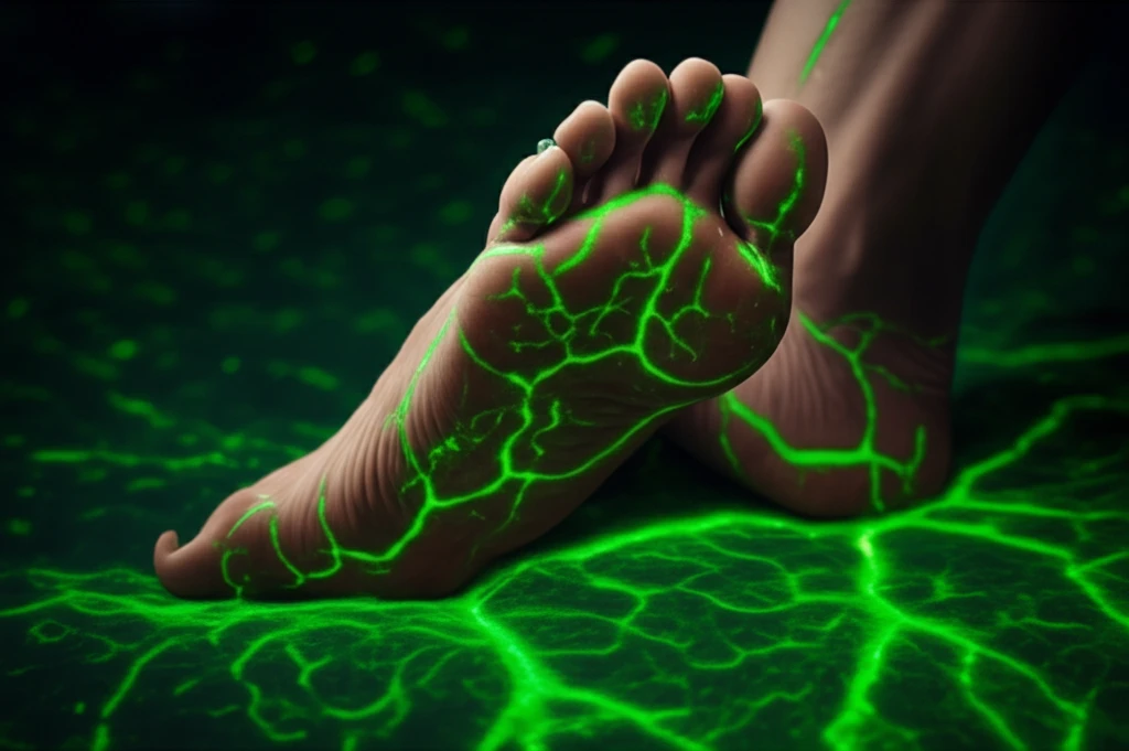
At its core, IG-FI leverages the unique properties of indocyanine green (ICG), a fluorescent dye that emits light when stimulated by near-infrared light. This allows clinicians to visualize blood flow in real-time. Here's how it works:

The process begins with the injection of ICG into the patient's bloodstream. As the dye circulates, it illuminates the vasculature, highlighting areas of adequate and compromised perfusion. A specialized camera captures the fluorescence emitted by the ICG, generating detailed images of blood flow dynamics.

  • Real-Time Visualization: IG-FI provides a dynamic view of blood flow, allowing for immediate assessment of perfusion.
  • Enhanced Precision: The fluorescent properties of ICG offer superior contrast, making it easier to identify areas of ischemia.
  • Non-Invasive Assessment: Unlike traditional angiography, IG-FI is non-invasive, reducing the risk of complications for patients.
The study's methodology involved a prospective analysis of 72 patients with critical limb ischemia undergoing surgical or endovascular revascularization. Researchers used IG-FI to measure perfusion in the foot and wound area before and after the intervention. They analyzed key parameters such as the curve of intensity, maximum intensity, and the increase in intensity to gauge the effectiveness of the revascularization.

A Promising Future for Ischemic Foot Treatment

IG-FI represents a significant advancement in the management of ischemic foot. By providing clinicians with a more precise and dynamic assessment of perfusion, this technology empowers them to make informed decisions, optimize treatment strategies, and ultimately, improve patient outcomes. As research continues to expand the applications of IG-FI, the future holds immense promise for individuals at risk of limb loss due to ischemia.

About this Article -

This article was crafted using a human-AI hybrid and collaborative approach. AI assisted our team with initial drafting, research insights, identifying key questions, and image generation. Our human editors guided topic selection, defined the angle, structured the content, ensured factual accuracy and relevance, refined the tone, and conducted thorough editing to deliver helpful, high-quality information.See our About page for more information.

This article is based on research published under:

DOI-LINK: 10.1016/j.avsg.2018.08.046, Alternate LINK

Title: Outcomes In 90 Patients Operated For A Venous Thoracic Outlet Syndrome

Subject: Cardiology and Cardiovascular Medicine

Journal: Annals of Vascular Surgery

Publisher: Elsevier BV

Authors: Jihéne Fendri, Olivier Coffin, Lucie Cameliere, Etienne Joguet, Claire Boirat, Laura Palcau, Ludovic Berger

Published: 2018-11-01

Everything You Need To Know

1

How does indocyanine green fluorescence imaging (IG-FI) allow doctors to 'see' blood flow in ischemic feet?

Indocyanine green fluorescence imaging (IG-FI) utilizes a fluorescent dye called indocyanine green (ICG). When ICG is injected into the bloodstream and stimulated by near-infrared light, it emits light, allowing clinicians to visualize blood flow in real-time. A specialized camera captures this fluorescence, creating detailed images of blood flow dynamics, revealing areas of both adequate and compromised perfusion.

2

What are the key advantages of using indocyanine green fluorescence imaging (IG-FI) over traditional methods for assessing ischemic foot?

Indocyanine green fluorescence imaging (IG-FI) offers real-time visualization of blood flow, enhanced precision in identifying areas of ischemia due to the fluorescent properties of indocyanine green (ICG), and it's a non-invasive assessment, reducing risks compared to traditional angiography. These benefits allow for better-informed decisions in treatment strategies.

3

What specific measurements were taken using indocyanine green fluorescence imaging (IG-FI) in the study involving patients with critical limb ischemia?

In the study presented, researchers used indocyanine green fluorescence imaging (IG-FI) to measure perfusion in the foot and wound area of 72 patients with critical limb ischemia before and after surgical or endovascular revascularization. They analyzed parameters like the curve of intensity, maximum intensity, and the increase in intensity to assess the effectiveness of the revascularization procedure.

4

How might indocyanine green fluorescence imaging (IG-FI) change how doctors treat ischemic foot, potentially reducing limb loss?

Indocyanine green fluorescence imaging (IG-FI) enhances clinical decision-making by providing a more precise and dynamic assessment of perfusion. This allows clinicians to optimize treatment strategies for ischemic foot, potentially improving patient outcomes and reducing the risk of limb loss. The ability to visualize blood flow in real-time is invaluable for guiding interventions.

5

What aspects of indocyanine green fluorescence imaging (IG-FI) for ischemic foot treatment still require further investigation and what are the broader implications?

While the study highlights the benefits of indocyanine green fluorescence imaging (IG-FI) in assessing perfusion for ischemic foot, it doesn't delve into the long-term outcomes of patients treated using IG-FI-guided interventions. Further research could explore the durability of improved perfusion, the impact on wound healing rates, and the overall long-term quality of life for patients who undergo IG-FI-guided treatment for ischemic foot. It would be beneficial to study the cost-effectiveness, and how it compares with traditional methods of angiography in managing ischemic foot.

Newsletter Subscribe

Subscribe to get the latest articles and insights directly in your inbox.