Illustration of a foot with weight-bearing scales and radiographic light, representing foot health and medical precision.

Unlocking Foot Health: How Weight-Bearing Radiographs Can Improve Your Diagnosis

"Understanding the Influence of Percentage Weight-Bearing on Foot Radiographs for Better Clinical Decisions"


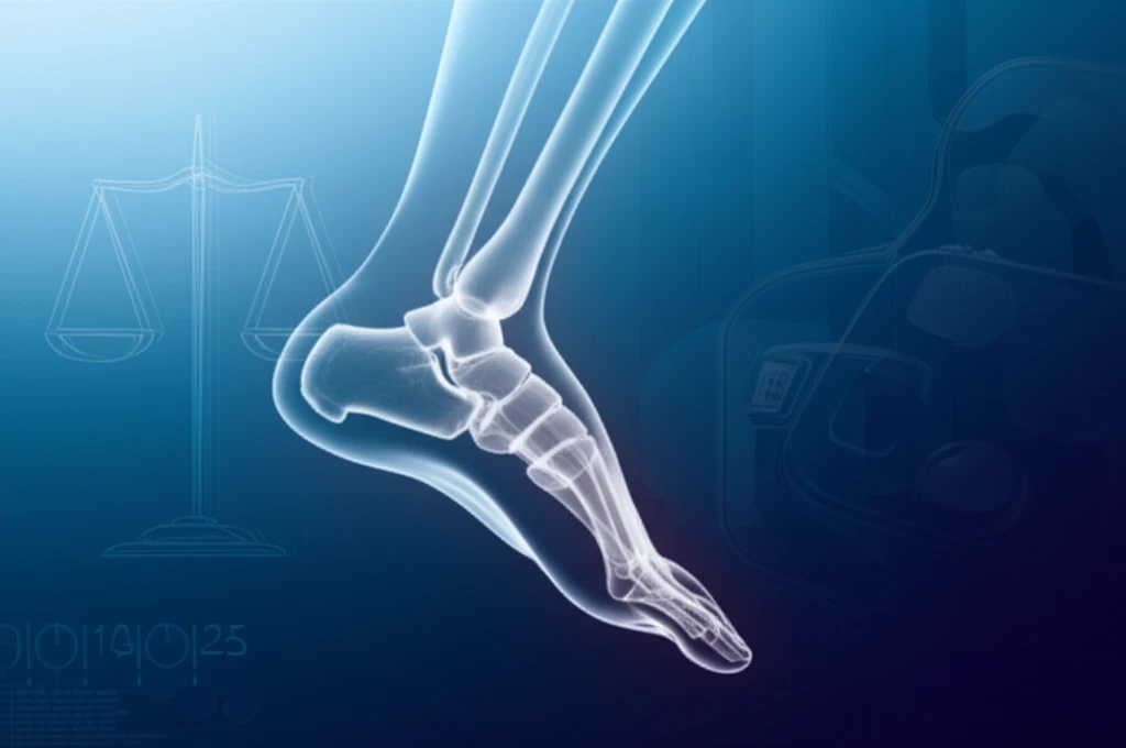
Foot radiographs are a cornerstone of diagnosing and managing various foot conditions. When a patient presents with foot pain or a suspected injury, one of the first steps a healthcare provider often takes is to order weight-bearing radiographs. These images, taken while the patient is standing, provide critical information about the foot's structure and alignment under the stresses of everyday activity.

Clinical decisions, from conservative treatments to surgical interventions, heavily rely on the insights gained from these weight-bearing radiographs. However, a crucial question remains: how do different levels of weight-bearing influence the measurements and interpretations derived from these images? Understanding this influence is essential for healthcare providers to make accurate and informed decisions.

Recent research has shed light on this very topic, investigating how varying percentages of weight-bearing affect specific radiographic measurements of the foot. This article delves into the findings of this study, exploring the clinical implications and offering practical guidance for leveraging weight-bearing radiographs to enhance foot health diagnoses.

Why Percentage of Weight-Bearing Matters: Unveiling the Research

Illustration of a foot with weight-bearing scales and radiographic light, representing foot health and medical precision.

A prospective study involving twenty healthy individuals examined foot radiographs under five different weight-bearing conditions: non-weight-bearing, 10% body weight, 25% body weight, 50% body weight, and 100% body weight. Researchers meticulously measured several key radiographic parameters under each condition, including:

Hallux valgus angle (HVA): The angle between the long axis of the first metatarsal and the first proximal phalanx, crucial for assessing bunions.

  • 1-2 intermetatarsal angle (IMA): The angle between the long axes of the first and second metatarsals, essential for evaluating forefoot deformities.
  • Talonavicular coverage angle (TNCA): The angle between the articular surfaces of the talus and navicular, indicating midfoot alignment.
  • Talocalcaneal angle (TCA): Also known as Kite's angle, this measurement reflects hindfoot alignment.
  • Forefoot width: Measured as the widest point of the metatarsal heads.
  • Lisfranc distance: The distance between the middle cuneiform and the second metatarsal, vital for assessing midfoot stability.
  • Cuboid height to ground (CHG): Perpendicular distance from the most inferior aspect of the cuboid to the horizontal supporting surface, reflecting arch height.
  • Talo-first metatarsal angle (TMA): Meary's angle, formed between the long axis of the talus and the first metatarsal, indicating overall foot alignment.
By comparing these measurements across the different weight-bearing conditions, the study aimed to determine how varying levels of weight-bearing influence radiographic measurements in the foot. The statistical analysis focused on identifying significant differences in each measurement across the different weight-bearing conditions, providing valuable insights into the dynamic changes occurring in the foot under load.

Turning Insights into Action: Optimizing Your Approach to Radiographs

The key takeaway is that the amount of weight placed on the foot can influence specific radiographic measurements. This means clinicians should be mindful of a patient's weight-bearing status when interpreting radiographs. Encouraging patients to bear at least 25% of their body weight can provide a more accurate representation of foot alignment, comparable to a full weight-bearing stance. For graduated post-injury or postoperative weight-bearing regimens, understanding these thresholds can refine treatment protocols, ensuring that patients receive the most appropriate and effective care based on reliable imaging.

About this Article -

This article was crafted using a human-AI hybrid and collaborative approach. AI assisted our team with initial drafting, research insights, identifying key questions, and image generation. Our human editors guided topic selection, defined the angle, structured the content, ensured factual accuracy and relevance, refined the tone, and conducted thorough editing to deliver helpful, high-quality information.See our About page for more information.

Everything You Need To Know

1

What are weight-bearing radiographs, and why are they important for diagnosing foot problems?

Weight-bearing radiographs are X-ray images taken while a patient is standing. These radiographs are significant because they provide essential information about the foot's structure and alignment under the stresses of everyday activity. Unlike non-weight-bearing radiographs, weight-bearing images allow healthcare providers to assess how the foot functions under load, which is crucial for diagnosing and managing various foot conditions. The information obtained from these images directly influences clinical decisions, ranging from conservative treatments to surgical interventions, making them a cornerstone in foot health management.

2

Why is the percentage of weight-bearing important when taking foot radiographs?

The percentage of weight-bearing during a radiograph matters because different levels of weight-bearing can influence the measurements and interpretations derived from the images. Research has shown that varying percentages of weight-bearing affect specific radiographic measurements of the foot, such as the hallux valgus angle (HVA), 1-2 intermetatarsal angle (IMA), talonavicular coverage angle (TNCA), talocalcaneal angle (TCA), forefoot width, Lisfranc distance, cuboid height to ground (CHG), and talo-first metatarsal angle (TMA). Understanding this influence is essential for healthcare providers to make accurate and informed decisions, especially when patients are on graduated weight-bearing regimens.

3

What specific measurements are taken on foot radiographs, and what do they tell us?

Several key radiographic parameters are measured to assess foot alignment and conditions. These include: * Hallux valgus angle (HVA): Assesses bunions. * 1-2 intermetatarsal angle (IMA): Evaluates forefoot deformities. * Talonavicular coverage angle (TNCA): Indicates midfoot alignment. * Talocalcaneal angle (TCA): Reflects hindfoot alignment. * Forefoot width: Measured at the widest point of the metatarsal heads. * Lisfranc distance: Assesses midfoot stability. * Cuboid height to ground (CHG): Reflects arch height. * Talo-first metatarsal angle (TMA): Indicates overall foot alignment. These measurements are critical for diagnosing a range of foot conditions and guiding treatment decisions.

4

What percentage of weight-bearing is considered sufficient for an accurate foot radiograph?

According to research, encouraging patients to bear at least 25% of their body weight during a radiograph can provide a more accurate representation of foot alignment, comparable to a full weight-bearing stance. This is significant because it helps ensure that the radiographic measurements reflect how the foot functions under a more realistic load. For patients on graduated post-injury or postoperative weight-bearing regimens, understanding these thresholds can refine treatment protocols and ensure they receive the most appropriate and effective care based on reliable imaging.

5

Does the amount of weight placed on the foot during a radiograph actually change the measurements?

Yes, the amount of weight placed on the foot during a radiograph can indeed influence the specific radiographic measurements obtained. This means clinicians should be mindful of a patient's weight-bearing status when interpreting radiographs. Failure to consider the weight-bearing status may lead to inaccurate assessments of foot alignment and potentially inappropriate treatment decisions. Therefore, it is crucial to ensure that the weight-bearing status is documented and considered when analyzing radiographic images.

Newsletter Subscribe

Subscribe to get the latest articles and insights directly in your inbox.