Unlocking Epilepsy: How Brain Tissue Analysis is Paving the Way for New Treatments
"Proteomic analysis of epileptic neocortex reveals potential targets for managing seizures and improving patient outcomes."
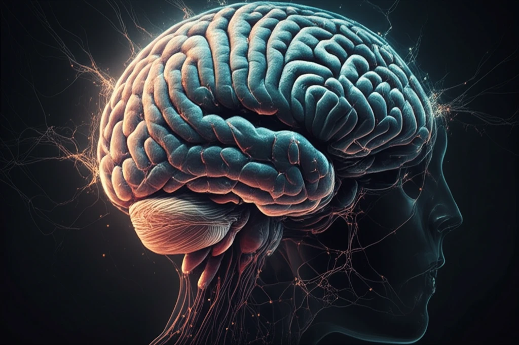
Epilepsy, a neurological disorder affecting millions worldwide, remains a significant challenge for both patients and medical professionals. Characterized by recurrent seizures, epilepsy's underlying molecular mechanisms are still not fully understood, making effective treatment elusive for many.
For those who don't respond to medication, surgical removal of brain regions that trigger seizures can offer relief. This procedure provides an invaluable opportunity to study human brain tissue and understand the cellular and molecular basis of epilepsy. A recent study delved into this area, seeking to identify common proteomic patterns in brain regions responsible for generating epileptic discharges.
This article explores the findings of this proteomic study, which analyzed neocortical tissue from six patients with refractory epilepsy. The research aimed to uncover how protein expression differs between regions that produce epileptic discharges and those that do not, offering new insights into potential therapeutic targets.
Decoding the Epileptic Brain: A Proteomic Approach
The research team conducted a detailed proteomic analysis as part of the Systems Biology of Epilepsy Project (SBEP). They studied in vivo electrophysiologically characterized human brain samples taken from patients undergoing surgery for refractory epilepsy. The study's unique design involved comparing protein expression within the same patient—contrasting highly epileptic regions with less epileptic ones.
- Interictal Spikes: Electrical activity between seizures, serving as a key indicator.
- 2D-DIGE Analysis: Identifying 31 protein spots with notable expression changes.
- GFAP Findings: Glial fibrillary acidic protein consistently downregulated in high-spiking brain tissue.
Implications and Future Directions
This study’s innovative approach, combined with proteomic data analysis, predicts new glial changes, increased angiogenesis, and alterations in cytoskeleton and neuronal projections in regions with high interictal spiking. Validating these findings through quantitative histological staining of the same tissues confirmed vascular and glial changes, providing new insights into neocortical epilepsy's structural and functional underpinnings. These discoveries open new avenues for targeted treatments and interventions, offering hope for more effective management of this complex neurological disorder.