3D jaw model showing teeth development stages using CBCT technology

Unlocking Dental Secrets: How 3D Scans Revolutionize Tooth Development Analysis

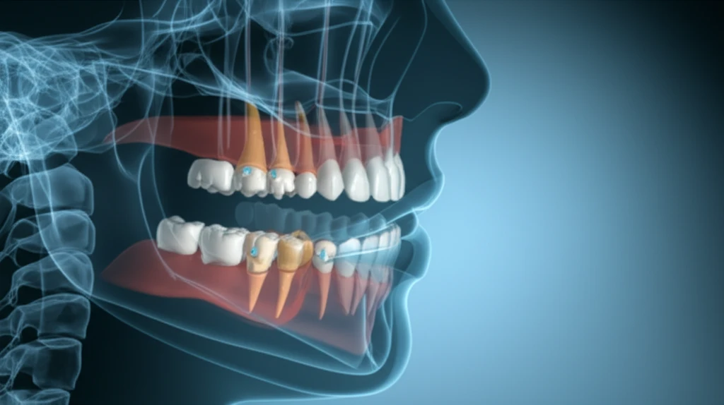
"Cone-beam computed tomography (CBCT) offers unprecedented insight into the hidden world of tooth development, paving the way for more accurate diagnoses and treatment plans."


Understanding how permanent teeth develop is crucial for effective dental care, influencing diagnosis, treatment strategies, and overall outcomes. Traditionally, dental professionals have relied on two-dimensional X-ray images to assess tooth development. However, these images only provide a limited view of complex three-dimensional structures.

Now, advancements in imaging technology, particularly cone-beam computed tomography (CBCT), are changing the game. CBCT delivers detailed, high-resolution, three-dimensional images of the oral structures, allowing for earlier and more precise detection of any abnormalities.

A recent study published in the Dental Press Journal of Orthodontics investigates the use of CBCT for measuring tooth development. The study aims to determine the standard linear measurements of permanent teeth at various developmental stages, providing a valuable resource for dental professionals.

CBCT: A New Dimension in Dental Development Assessment

3D jaw model showing teeth development stages using CBCT technology

The study leverages CBCT technology to analyze the development of permanent teeth in 18 patients aged 3 to 20. CBCT images were acquired using the i-CAT system, and measurements were taken using the i-CAT software. The study focused on analyzing 238 teeth at varying development stages in both coronal and sagittal planes.

Researchers established a method based on specific anatomical landmarks related to crown and root development. By measuring the distances between these landmarks, a quantitative model was created to assess the initial and final development stages for all dental groups.

Here’s a breakdown of the key measurements:
The measurements were made specifically for each root. The B'C' reference for teeth with more than one root used the mean distance between roots. Using these measurements a quantitative model with five scores was suggested for all dental groups (with the exception of the third molar): 0 = absence of dental crypt; 1 = presence of dental crypt; 2 = dental crown partially formed; 3 = dental crown completely formed; 4 = beginning of root formation - open apex; 5 = end of root formation - closed apex) (Fig 1).

Implications for Diagnosis and Treatment

The study's findings align with previous research, confirming the reliability of CBCT for assessing tooth development. The measurements of dental crowns and roots acquired through CBCT imaging have significant clinical and research applications, providing a non-invasive method for in vivo studies.

CBCT images can significantly improve diagnosis, treatment planning, and outcome prediction in various dental specialties. The three-dimensional visualization allows for a more comprehensive understanding of tooth structures and potential abnormalities.

While the study provides valuable insights, the authors recommend further research to minimize methodological variables and refine the CBCT imaging process for even greater accuracy.

About this Article -

This article was crafted using a human-AI hybrid and collaborative approach. AI assisted our team with initial drafting, research insights, identifying key questions, and image generation. Our human editors guided topic selection, defined the angle, structured the content, ensured factual accuracy and relevance, refined the tone, and conducted thorough editing to deliver helpful, high-quality information.See our About page for more information.

Everything You Need To Know

1

What is cone-beam computed tomography (CBCT), and how does it work?

Cone-beam computed tomography (CBCT) is a type of imaging technology that provides three-dimensional (3D) images of the teeth and surrounding oral structures. Unlike traditional two-dimensional X-rays, CBCT offers a more detailed and comprehensive view, allowing dental professionals to assess tooth development and identify abnormalities more accurately. The imaging utilizes the i-CAT system to acquire the images for analysis.

2

Why is cone-beam computed tomography (CBCT) so important in the context of dental development analysis?

The significance of CBCT lies in its ability to revolutionize dental development analysis. Compared to traditional methods, such as two-dimensional X-rays which provide limited views, CBCT allows for a non-invasive, detailed examination of tooth development stages. This technology facilitates early detection of potential issues, thereby improving diagnosis and treatment planning. The high-resolution 3D images help dental professionals make informed decisions, leading to better patient outcomes.

3

How does the study measure tooth development using cone-beam computed tomography (CBCT) and anatomical landmarks?

The study uses specific anatomical landmarks to measure the distances between crown and root development. Using these measurements, a quantitative model was created to assess the initial and final development stages for all dental groups. The measurements were made specifically for each root. The B'C' reference for teeth with more than one root used the mean distance between roots. This method is designed to standardize the assessment of tooth development and provides a reliable framework for clinical and research applications.

4

What are the implications of using cone-beam computed tomography (CBCT) for diagnosis and treatment in dentistry?

CBCT's use has profound implications for dental diagnosis and treatment. By providing detailed 3D images, CBCT allows for the early detection of abnormalities in tooth development. This can lead to more accurate diagnoses and the development of tailored treatment plans. Moreover, the non-invasive nature of CBCT means that it provides a method for *in vivo* studies, allowing researchers to study tooth development without invasive procedures. This can ultimately lead to improvements in dental care and improved patient outcomes.

5

What is the scoring system used in the quantitative model for assessing tooth development with cone-beam computed tomography (CBCT)?

The quantitative model, based on CBCT measurements, uses a scoring system (0-5) to classify the development stages of teeth. The scores range from the absence of the dental crypt (0) to the end of root formation (5). This system helps to standardize the assessment of tooth development stages, aiding in the early detection of potential issues. This method is applicable to all dental groups with the exception of the third molar.

Newsletter Subscribe

Subscribe to get the latest articles and insights directly in your inbox.