Protein kinase enzyme with nucleotides bound to the active site.

Unlocking Cellular Secrets: How a Protein Kinase Reveals New Pathways in Cell Regulation

"Saccharomyces cerevisiae CK2α's multiple binding modes offer a fresh perspective on ADP/GDP release, challenging conventional models of protein kinase activity."


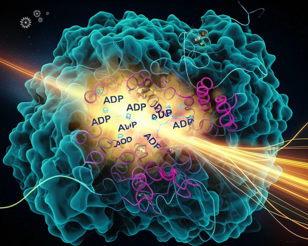
Protein kinases are fundamental to cell regulation, acting as master switches that control a myriad of processes. Among these, CK2 stands out as a highly conserved and constitutively active enzyme, crucial for everything from cell growth to programmed cell death. What makes CK2 particularly intriguing is its ability to use both ATP and GTP as energy sources, setting it apart from its kinase counterparts.

Recent research has delved into the intricate workings of Saccharomyces cerevisiae CK2α (scCK2α), a catalytic subunit of CK2 in yeast. By examining its crystal structures in complex with various nucleotide analogues and divalent cations, scientists have uncovered novel binding modes that hint at a unique mechanism for releasing ADP/GDP, the byproducts of its activity. This discovery challenges existing models and opens new avenues for understanding kinase function.

This article explores the groundbreaking findings regarding scCK2α's structure and function, translating complex research into accessible insights. We will unravel how scCK2α's distinctive features provide clues about its co-substrate hydrolysis product release pathway, its dual co-substrate specificity, and the role of a unique insertion region in maintaining its constitutively active state.

Decoding the Structure: Unique Features of scCK2α

Protein kinase enzyme with nucleotides bound to the active site.

The crystal structures of scCK2α, complexed with GMPPNP, ATP, and AMPPN in the presence of magnesium or manganese ions, reveal a high degree of similarity to other known CK2 structures. Like its counterparts, scCK2α comprises two domains with a co-substrate nestled in the cleft between them. However, scCK2α exhibits three key distinctions:

First, interactions between Lys45-Glu53 and Arg48-Glu53 cause Lys50 to adopt a unique conformation that stabilizes the y-phosphate of the co-substrate. This lessens the necessity for the 'essential divalent cation' typically required for activity.

  • Multiple Binding Modes: scCK2α exhibits multiple nucleotide-divalent cation binding modes in its active site, differentiating it from the two-divalent-cation-occupied active sites of Zea mays CK2α and human CK2α.
  • Conformational Changes: A conformational shift in Glu53 within the scCK2α-AMPPN complex disrupts its interaction with Lys45 and Arg48, resulting in a more open co-substrate binding pocket.
  • ADP/GDP Release Pathway: The open pocket suggests a potential pathway for ADP/GDP release. However, the NE1 atom of the Trp residue in the 'DWG motif' forms a hydrogen bond to the O atom of Leu212, potentially hindering the 'DFG-in flip to DFG-out' model commonly found in eukaryotic protein kinases.
The unique insertion region within scCK2α further sets it apart. This region contributes to maintaining the constitutively active conformation of the catalytic site, without engaging in interactions with regulatory subunits. These structural nuances suggest that scCK2α operates via a mechanism distinct from its homologues.

Implications and Future Directions

The discovery of multiple nucleotide-divalent cation binding modes in scCK2α, coupled with the proposed ADP/GDP release pathway, challenges our established understanding of protein kinase function. The research highlights the diverse mechanisms employed by enzymes to regulate cellular processes.

Further investigation into the conformational changes observed in scCK2α, particularly the role of the insertion region and the interactions of key residues like Lys50, Glu53, Arg161, and Lys197, promises to reveal more about the intricate regulation of CK2 activity.

By illuminating the unique characteristics of scCK2α, this research paves the way for developing targeted therapies that can modulate CK2 activity in various diseases, offering hope for innovative treatment strategies.

About this Article -

This article was crafted using a human-AI hybrid and collaborative approach. AI assisted our team with initial drafting, research insights, identifying key questions, and image generation. Our human editors guided topic selection, defined the angle, structured the content, ensured factual accuracy and relevance, refined the tone, and conducted thorough editing to deliver helpful, high-quality information.See our About page for more information.

This article is based on research published under:

DOI-LINK: 10.1107/s1399004713027879, Alternate LINK

Title: The Multiple Nucleotide–Divalent Cation Binding Modes OfSaccharomyces CerevisiaeCk2Α Indicate A Possible Co-Substrate Hydrolysis Product (Adp/Gdp) Release Pathway

Subject: General Medicine

Journal: Acta Crystallographica Section D Biological Crystallography

Publisher: International Union of Crystallography (IUCr)

Authors: Huihui Liu, Hong Wang, Maikun Teng, Xu Li

Published: 2014-01-30

Everything You Need To Know

1

What is the role of protein kinases in cell regulation?

Protein kinases are essential regulators within cells, functioning like master switches that govern a variety of processes. The research focuses on Saccharomyces cerevisiae CK2α (scCK2α), a specific type of protein kinase. It highlights the significance of understanding these kinases because they are fundamental to cellular operations, encompassing everything from growth to programmed cell death. ScCK2α is particularly intriguing because it can utilize both ATP and GTP, distinguishing it from other kinases.

2

What makes the multiple binding modes of scCK2α significant?

The multiple binding modes of scCK2α are significant because they challenge established models of how protein kinases function. The study reveals that scCK2α, unlike other CK2 structures, exhibits multiple nucleotide-divalent cation binding modes. This uniqueness suggests a different mechanism for releasing ADP/GDP, the products of its catalytic activity. This insight is crucial as it redefines our understanding of cellular regulation and opens new pathways for investigating kinase function.

3

How does scCK2α release ADP/GDP?

The ADP/GDP release pathway is the mechanism by which scCK2α releases ADP/GDP after a reaction. The study suggests that scCK2α might have a unique pathway for releasing these byproducts. This pathway is hinted at by the open co-substrate binding pocket observed in scCK2α complexes. However, the NE1 atom of the Trp residue in the 'DWG motif' forms a hydrogen bond to the O atom of Leu212, which potentially hinders the 'DFG-in flip to DFG-out' model commonly found in eukaryotic protein kinases.

4

Why is the unique insertion region in scCK2α important?

The unique insertion region in scCK2α is a distinctive feature that contributes to maintaining its constitutively active state. This region helps keep the catalytic site in a conformation that is always active, without requiring interactions with regulatory subunits. This feature contrasts with other kinases and suggests that scCK2α operates through a mechanism distinct from its counterparts. The implications are that this unique region is essential for its consistent activity, making it a key component in cellular regulation.

5

What specific aspects of scCK2α are being studied, and why?

The study focuses on scCK2α by examining its structure and function, specifically how it binds with nucleotide analogues and divalent cations. By studying the crystal structures of scCK2α complexed with various molecules like GMPPNP, ATP, and AMPPN, scientists could identify novel binding modes. This research offers insights into the mechanisms of cellular regulation, particularly in the context of yeast. Understanding scCK2α helps researchers to better understand the fundamental processes governing cell behavior and has the potential to inform future studies on protein kinase function.

Newsletter Subscribe

Subscribe to get the latest articles and insights directly in your inbox.