Microscopic view of fungal cells stained with fluorescent dyes, illustrating apoptosis.

Unlocking Cell Secrets: How a Simple Stain Reveals the Mysteries of Cell Death

"Discover how scientists are using everyday dyes to explore apoptosis, the key to understanding diseases like cancer and Alzheimer's."


Apoptosis, or programmed cell death, is a fundamental process that our bodies use to maintain health. Think of it as the body's way of tidying up, getting rid of cells that are damaged, old, or no longer needed. This process is crucial for everything from embryonic development to preventing cancer. Scientists are constantly working to understand exactly how apoptosis works, because when it goes wrong, it can lead to serious diseases.

One of the ongoing challenges in cell biology is accurately detecting and studying apoptosis in action. Traditional methods can be complex and time-consuming, often requiring specialized equipment and extensive training. Researchers need simpler, faster, and more reliable ways to observe and analyze this critical process in living cells.

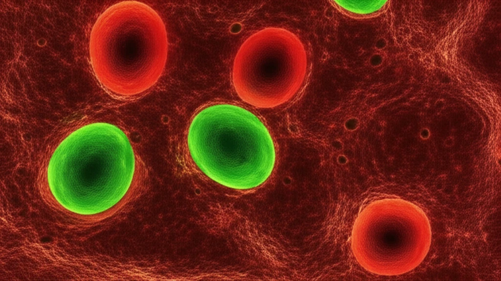
Now, imagine being able to peek into the microscopic world of cells and instantly identify which ones are undergoing apoptosis. That’s the promise of a technique using common dyes: ethidium bromide and acridine orange. This method offers a straightforward way to visualize and differentiate between healthy, apoptotic, and necrotic cells, providing valuable insights into cellular processes and disease mechanisms.

Why Use Simple Stains to Uncover Complex Cell Processes?

Microscopic view of fungal cells stained with fluorescent dyes, illustrating apoptosis.

For years, scientists have relied on sophisticated techniques to study apoptosis. However, these methods often have limitations. Some require multiple steps, making them time-intensive and increasing the risk of damaging the cells. Others struggle to distinguish between different types of cell death, such as apoptosis and necrosis, leading to potential inaccuracies.

That's where ethidium bromide (EB) and acridine orange (AO) come in. These fluorescent dyes offer a simple yet powerful way to visualize cell death. AO penetrates all cells, staining the nuclei green and highlighting the structure of the chromatin. EB, on the other hand, can only enter cells with damaged membranes, staining them red-orange. This allows researchers to easily differentiate between healthy cells (green nuclei), early apoptotic cells (condensed green nuclei), and late apoptotic or necrotic cells (red-orange nuclei).

  • Simplicity and Speed: The EB/AO staining method is quick and easy to perform, requiring minimal equipment and training.
  • Clear Differentiation: The distinct colors allow for easy identification of cells in different stages of cell death.
  • Versatility: This technique can be used on various cell types and organisms, making it a valuable tool for diverse research applications.
In a recent study, researchers used EB and AO to investigate apoptosis in Aspergillus nidulans, a type of fungus commonly used in genetic research. By staining micro-colonies of the fungus with these dyes, they were able to observe the effects of ultraviolet (UV) light on cell death. The results showed that UV radiation induced apoptosis in the fungal cells, demonstrating the effectiveness of the EB/AO staining method for studying cell death in this organism.

The Future of Cell Death Research: Simple Tools, Big Discoveries

The EB/AO staining technique offers a valuable tool for researchers studying apoptosis and other forms of cell death. Its simplicity, speed, and versatility make it an attractive alternative to more complex methods, allowing scientists to gain new insights into the fundamental processes that govern cell life and death. As research continues, expect to see even wider applications of this technique, paving the way for new discoveries in medicine, biology, and beyond.

About this Article -

This article was crafted using a human-AI hybrid and collaborative approach. AI assisted our team with initial drafting, research insights, identifying key questions, and image generation. Our human editors guided topic selection, defined the angle, structured the content, ensured factual accuracy and relevance, refined the tone, and conducted thorough editing to deliver helpful, high-quality information.See our About page for more information.

This article is based on research published under:

DOI-LINK: 10.4238/2013.august.12.5, Alternate LINK

Title: In Situ Analysis Of Apoptosis In Aspergillus Nidulans With Ethidium Bromide And Acridine Orange

Subject: Genetics

Journal: Genetics and Molecular Research

Publisher: Genetics and Molecular Research

Authors: S.M. Leles, R. Factori, C.L.M.S.C. Rocha

Published: 2013-01-01

Everything You Need To Know

1

What is Apoptosis and why is it important?

Apoptosis, or programmed cell death, is a vital process where the body eliminates cells that are damaged, aged, or no longer needed. This is essential for healthy development, preventing diseases like cancer, and maintaining overall cellular balance. When Apoptosis goes wrong, it can lead to various health problems.

2

What is the main problem that Ethidium Bromide and Acridine Orange aim to solve?

The primary problem addressed by these methods is the need for simpler, faster, and more reliable techniques to observe and analyze Apoptosis. Traditional methods are often complex and time-consuming. The use of Ethidium Bromide (EB) and Acridine Orange (AO) offers a straightforward alternative to visualize and differentiate between healthy, apoptotic, and necrotic cells, enhancing the study of cellular processes and disease mechanisms.

3

How do Ethidium Bromide and Acridine Orange work to identify cell death?

Ethidium Bromide (EB) and Acridine Orange (AO) are used because of their ability to visualize cell death efficiently. AO penetrates all cells and stains the nuclei green. EB only enters cells with damaged membranes and stains them red-orange. This allows researchers to easily distinguish between healthy cells (green nuclei), early apoptotic cells (condensed green nuclei), and late apoptotic or necrotic cells (red-orange nuclei).

4

Why is the EB/AO staining method considered valuable?

The EB/AO staining method is significant due to its simplicity, speed, and versatility. It requires minimal equipment and training, allowing for quick and easy performance. The distinct colors provide clear differentiation between different stages of cell death. The technique's versatility makes it applicable across various cell types and organisms, offering a valuable tool for diverse research applications in medicine, biology, and beyond.

5

How was the EB/AO staining method applied in a specific study?

In a recent study, researchers utilized Ethidium Bromide (EB) and Acridine Orange (AO) to investigate Apoptosis in Aspergillus nidulans, a fungus used in genetic research. By staining micro-colonies with these dyes, the researchers observed the effects of ultraviolet (UV) light on cell death, proving the effectiveness of the EB/AO staining method in this context. This demonstrates how the staining method can be used to study the effects of external factors on cell death in various organisms.

Newsletter Subscribe

Subscribe to get the latest articles and insights directly in your inbox.