Surreal illustration of the microscopic world inside a bacterium.

Unlocking Bacterial Secrets: A New Way to See What's Inside

"Revolutionary X-ray technique reveals the hidden distribution of elements within bacteria, paving the way for breakthroughs in understanding cellular processes and fighting disease."


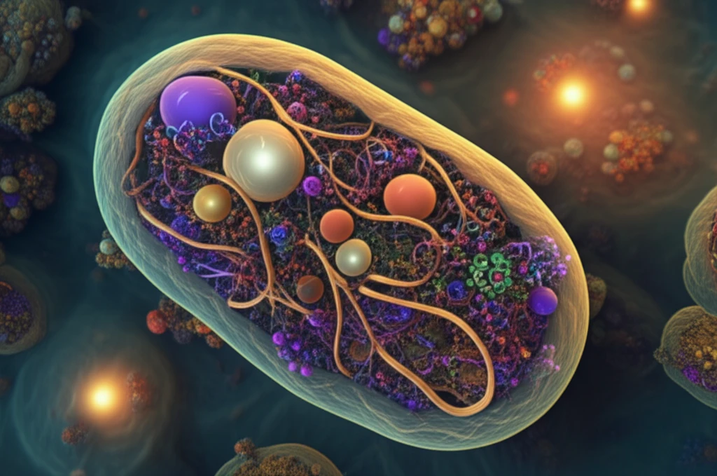
For years, scientists have been trying to get a clear picture of what's happening inside living cells. Trace elements like metals play crucial roles in cell function, but seeing exactly where they are and what they're doing has been a challenge. Traditional methods often require staining or sectioning cells, which can disrupt their natural state.

Now, a team of researchers has developed a powerful new approach using X-ray fluorescence (XRF) nanotomography. This technique allows them to create detailed three-dimensional images of the elemental distribution within single bacteria, all without the need for harsh treatments that could alter the cell's structure.

This article will explore how this innovative method works, what it has already revealed about the inner workings of bacteria, and what potential applications it holds for future research and medical advancements.

X-Ray Vision for the Microscopic World: How It Works

Surreal illustration of the microscopic world inside a bacterium.

The key to this breakthrough is the use of a focused X-ray beam, a mere 15 nanometers in size, at the Hard X-ray Nanoprobe beamline (HXN) at NSLS-II. This incredibly small beam allows scientists to scan across individual bacteria, mapping the location of different elements based on the fluorescent signals they emit when struck by the X-rays. This X-ray vision is achieved using specialized X-ray microscope located at a beamline with infrastructure for good vibration isolation and thermal stability

To preserve the bacteria's natural structure during imaging, the researchers embedded the cells in tiny sodium chloride crystals. This non-aqueous matrix helps maintain the cell's three-dimensional integrity while data is collected at room temperature. In addition, the X-ray setup allows for simultaneous ptychography, which provides structural information, so both chemical and structural data are collected at the same time.

  • X-ray Fluorescence (XRF): Maps the location of elements within the cell based on fluorescent signals.
  • Nanotomography: Creates a three-dimensional reconstruction of the cell's interior.
  • Ptychography: Provides structural information about the cell's components.
The beauty of this technique is that it doesn't require staining or sectioning the cells, which can introduce artifacts and alter their natural state. By combining XRF nanotomography and ptychography, researchers can get a complete picture of a cell's structure and chemical composition in its native environment.

What We've Learned & What's Next

Using this method, the researchers analyzed E. coli bacteria and discovered that calcium was evenly distributed throughout the cells, but zinc was not. Zinc was concentrated at the polar ends of the cells. This work provides insights into how essential elements are distributed in bacteria.

The ability to visualize the internal structure and chemical makeup of bacteria has implications for fighting disease, understanding the roles of essential elements, as well as bacterial behavior in different environment.

With further development, XRF nanotomography could become a routine tool for studying the inner workings of cells, leading to new discoveries in biology, medicine, and beyond. The insights gained could lead to more effective treatments for infections and a deeper understanding of life at the smallest scale.

About this Article -

This article was crafted using a human-AI hybrid and collaborative approach. AI assisted our team with initial drafting, research insights, identifying key questions, and image generation. Our human editors guided topic selection, defined the angle, structured the content, ensured factual accuracy and relevance, refined the tone, and conducted thorough editing to deliver helpful, high-quality information.See our About page for more information.

This article is based on research published under:

DOI-LINK: 10.1038/s41598-018-31461-y, Alternate LINK

Title: X-Ray Fluorescence Nanotomography Of Single Bacteria With A Sub-15 Nm Beam

Subject: Multidisciplinary

Journal: Scientific Reports

Publisher: Springer Science and Business Media LLC

Authors: Tiffany W. Victor, Lindsey M. Easthon, Mingyuan Ge, Katherine H. O’Toole, Randy J. Smith, Xiaojing Huang, Hanfei Yan, Karen N. Allen, Yong S. Chu, Lisa M. Miller

Published: 2018-09-07

Everything You Need To Know

1

What specific technique is used to image the internal structure of the bacteria?

The new X-ray technique uses X-ray Fluorescence (XRF) nanotomography. This method employs a focused X-ray beam, just 15 nanometers in size, to scan individual bacteria. It maps the location of different elements within the bacteria based on the fluorescent signals they emit when struck by the X-rays. In addition to XRF, the technique also uses ptychography to gather structural information. The combination of these methods provides a complete picture of a cell's structure and chemical composition.

2

Why is the use of X-ray Fluorescence (XRF) nanotomography important?

X-ray Fluorescence (XRF) nanotomography is essential because it allows scientists to visualize the internal structure and chemical makeup of bacteria without damaging them. Traditional methods often involve staining or sectioning cells, which can disrupt their natural state and introduce artifacts. This new technique provides a more accurate view of the bacteria's natural state, opening doors to understanding cellular processes and fighting disease. This is important as trace elements, like metals, play crucial roles in cell function, and knowing their exact location is vital.

3

What is the role of the Hard X-ray Nanoprobe beamline (HXN) at NSLS-II?

The Hard X-ray Nanoprobe beamline (HXN) at NSLS-II is critical because it provides the necessary infrastructure for this groundbreaking research. It houses a specialized X-ray microscope and is equipped with features like good vibration isolation and thermal stability. These features are essential for generating the highly focused X-ray beam, just 15 nanometers in size, used in X-ray Fluorescence (XRF) nanotomography. Without the HXN, this type of detailed imaging of bacteria would not be possible.

4

What were the key findings from the analysis of E. coli bacteria?

The researchers analyzed E. coli bacteria using this technique and discovered that calcium was evenly distributed throughout the cells, but zinc was not. Zinc was concentrated at the polar ends of the cells. This is significant because it provides insights into how essential elements are distributed in bacteria. Understanding the distribution of these elements can help scientists understand the function of the bacteria. This could eventually help to identify potential drug targets or understand how bacteria cause infections.

5

How does ptychography contribute to this new imaging method?

Ptychography is used to provide structural information about the cell's components. This is achieved simultaneously with the X-ray Fluorescence (XRF) nanotomography, which maps the elemental distribution. Combining these two methods allows researchers to obtain a complete picture of a cell's structure and chemical composition. This is important because it allows for a more comprehensive understanding of the bacteria, enabling a more detailed study of how it functions, and how to combat infections.

Newsletter Subscribe

Subscribe to get the latest articles and insights directly in your inbox.