Cellular pathways leading to a stylized eye, symbolizing precise protein targeting for vision.

Unlock Your Vision: How Cells Target Key Proteins to the Right Place

"Scientists discover an important signaling system that ensures vital components reach their destinations in eye cells, paving the way for a deeper understanding of visual health."


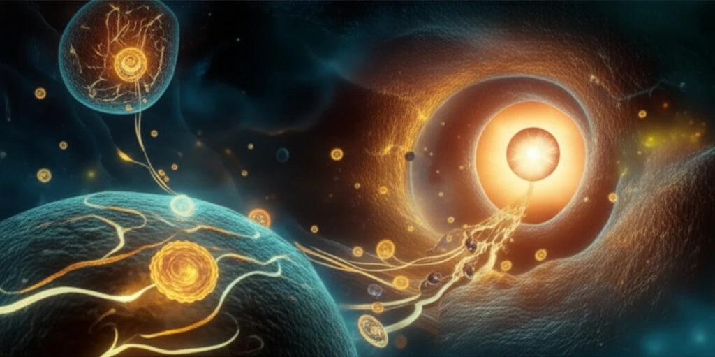
Our vision depends on a complex orchestra of cellular processes, with individual cells performing specialized roles to ensure we perceive the world around us accurately. Among these cells, photoreceptors in the retina are critical. They transform light into electrical signals that the brain interprets as images. The proper function of photoreceptors relies on having specific proteins in the right locations.

One such protein is the HCN1 channel, a vital component found in the inner segment of photoreceptors. HCN1 acts like a gatekeeper, regulating electrical signals that shape our vision, especially in varying light conditions. It's not enough for HCN1 to simply exist within the cell; it must be precisely delivered to its designated spot to do its job effectively. If HCN1 is mislocalized, visual processing can be impaired.

Scientists have long been puzzled by how cells manage to get proteins like HCN1 to their correct locations. It's a bit like navigating a complex city, where each protein needs to find its specific address. New research from the University of Iowa has uncovered a key piece of this puzzle, identifying a sophisticated 'addressing' system that guides HCN1 channels to their proper place within photoreceptor cells.

Decoding the Cellular GPS: How HCN1 Finds Its Way

Cellular pathways leading to a stylized eye, symbolizing precise protein targeting for vision.

The research pinpoints two critical signals that act in concert to direct HCN1 channels: an ER export signal and an ER retention signal. Think of these signals as opposing forces, each playing a role in ensuring the right balance of HCN1 at the cell surface. The ER (endoplasmic reticulum) is a network within the cell that acts as a manufacturing and distribution center. The ER export signal acts like a 'go' command, helping HCN1 channels leave the ER and begin their journey to the plasma membrane (the cell's outer boundary).

On the other hand, the ER retention signal functions like a 'wait' command, keeping HCN1 channels within the ER if they're not quite ready to be deployed. This retention signal ensures that only properly formed and functional HCN1 channels make it to the cell surface. The interplay between these two signals is crucial for maintaining the correct amount of HCN1 at the plasma membrane, ensuring optimal visual function.

  • ER Export Signal: Facilitates the movement of HCN1 channels out of the endoplasmic reticulum.
  • ER Retention Signal: Temporarily holds HCN1 channels within the ER, ensuring quality control.
  • Balance: The equilibrium between these signals dictates the amount of HCN1 present at the cell surface.
The research team identified a specific sequence of amino acids (the building blocks of proteins) within the HCN1 channel that acts as the ER export signal. This sequence, containing a leucine-based motif, essentially tells the cell, 'This HCN1 channel is ready for export.' By manipulating this signal in the lab, the scientists were able to alter the localization of HCN1 channels, demonstrating its importance in the targeting process. Further experiments revealed that this ER export signal overpowers the ER retention signal, ensuring that HCN1 reaches its destination. This delicate balance of export and retention guarantees that a sufficient amount of HCN1 is available at the cell surface to carry out its vital role in shaping our vision.

Why This Discovery Matters for Your Eye Health

This research is more than just an academic exercise; it has real implications for understanding and treating vision disorders. By deciphering the cellular mechanisms that govern protein localization, scientists can potentially develop therapies that target these pathways to correct imbalances. For example, if a disease process disrupts the ER export signal for HCN1, leading to its mislocalization, a future therapy might aim to boost that signal, restoring proper HCN1 function and improving vision. These findings open new avenues for research into a variety of vision-related conditions and could one day lead to innovative treatments that protect and enhance our sight.

About this Article -

This article was crafted using a human-AI hybrid and collaborative approach. AI assisted our team with initial drafting, research insights, identifying key questions, and image generation. Our human editors guided topic selection, defined the angle, structured the content, ensured factual accuracy and relevance, refined the tone, and conducted thorough editing to deliver helpful, high-quality information.See our About page for more information.

This article is based on research published under:

DOI-LINK: 10.1167/iovs.15-16902, Alternate LINK

Title: An N-Terminal Er Export Signal Facilitates The Plasma Membrane Targeting Of Hcn1 Channels In Photoreceptors

Subject: General Medicine

Journal: Investigative Opthalmology & Visual Science

Publisher: Association for Research in Vision and Ophthalmology (ARVO)

Authors: Yuan Pan, Joseph G. Laird, David M. Yamaguchi, Sheila A. Baker

Published: 2015-06-01

Everything You Need To Know

1

What is the role of HCN1 channels in photoreceptor cells?

HCN1 channels are vital components in the inner segment of photoreceptors. They act as gatekeepers, regulating electrical signals that shape our vision, especially in varying light conditions. It is crucial for HCN1 to be precisely delivered to its designated spot in order to function effectively, as mislocalization can impair visual processing. While the research focuses on the importance of HCN1 in photoreceptors, it does not delve into the specific types of light conditions it impacts, such as scotopic or photopic vision.

2

How do cells ensure that proteins like HCN1 reach their correct locations within the cell?

Cells use a sophisticated 'addressing' system to guide proteins like HCN1 to their proper place. For HCN1 channels, this involves two critical signals: an ER export signal and an ER retention signal. The ER export signal helps HCN1 channels leave the endoplasmic reticulum (ER) and begin their journey to the plasma membrane. The ER retention signal keeps HCN1 channels within the ER if they're not quite ready to be deployed, ensuring that only properly formed and functional HCN1 channels make it to the cell surface. The equilibrium between these signals dictates the amount of HCN1 present at the cell surface. The research does not specify how the ER identifies misfolded proteins. It only mentions that the ER retention signal ensures that only properly formed HCN1 makes it to the surface.

3

What are the ER export and ER retention signals, and how do they work together to target HCN1?

The ER export signal acts like a 'go' command, helping HCN1 channels leave the endoplasmic reticulum (ER) and begin their journey to the plasma membrane. It contains a specific sequence of amino acids, including a leucine-based motif. The ER retention signal functions like a 'wait' command, temporarily holding HCN1 channels within the ER if they're not yet ready to be deployed. The interplay between these two signals is crucial for maintaining the correct amount of HCN1 at the plasma membrane. The ER export signal overpowers the ER retention signal, ensuring that HCN1 reaches its destination. The research does not identify specific proteins that interact with the ER export and retention signals, only that the balance is important.

4

How might this research on HCN1 localization impact the treatment of vision disorders?

By deciphering the cellular mechanisms that govern protein localization, scientists can potentially develop therapies that target these pathways to correct imbalances that cause vision disorders. If a disease process disrupts the ER export signal for HCN1, leading to its mislocalization, a future therapy might aim to boost that signal, restoring proper HCN1 function and improving vision. This approach opens new avenues for research into various vision-related conditions. However, the research does not explicitly describe the steps in taking this knowledge to create a novel drug. It only hints at the potential for future therapies.

5

What is the significance of the balance between the ER export signal and the ER retention signal for HCN1 channels, and how does it affect vision?

The balance between the ER export signal and the ER retention signal is critical for ensuring that the correct amount of HCN1 is available at the cell surface. The ER export signal facilitates the movement of HCN1 channels out of the endoplasmic reticulum, while the ER retention signal temporarily holds HCN1 channels within the ER for quality control. The ER export signal overpowering the ER retention signal guarantees that a sufficient amount of HCN1 is present at the cell surface to carry out its role in shaping vision. Without this balance, there is a chance of incorrect protein localization and impaired vision. The research highlights the importance of this balance for visual function, without further explanation as to why this protein balance and movement is so important for vision.

Newsletter Subscribe

Subscribe to get the latest articles and insights directly in your inbox.