Surreal illustration of a healthy spine intertwined with glowing embryonic cells, symbolizing regeneration.

Unlock Your Spine's Secrets: How Embryonic Cells Could Revolutionize Back Pain Treatment

"Decoding the development of the spine's shock absorbers to pave the way for regenerative therapies."


Back pain, a relentless tormentor, plagues a staggering 85% of adults at some point in their lives. Beyond the personal suffering, the financial burden is immense, costing the United States over $100 billion annually. Current treatments, ranging from physical therapy and injections to spinal fusion and disc replacement, often provide temporary relief without addressing the root cause: intervertebral disc degeneration.

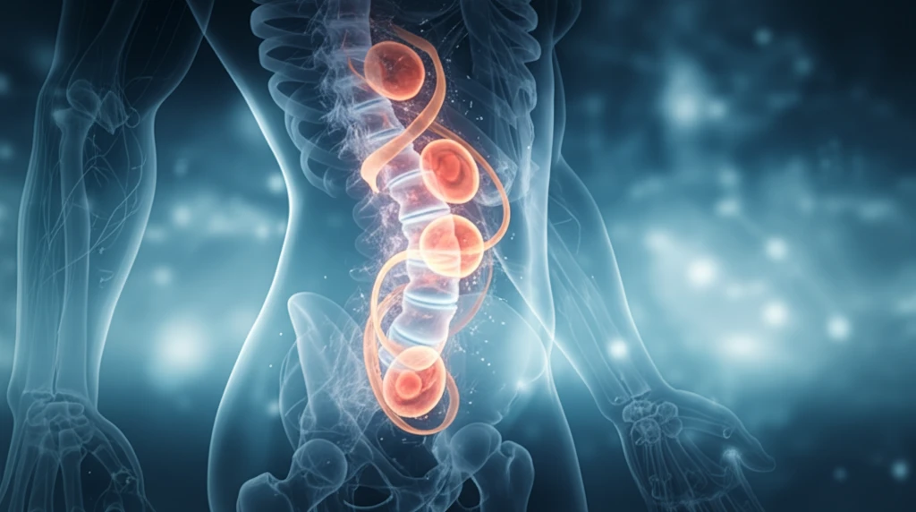
Intervertebral discs, the spine's unsung heroes, are complex structures comprised of the nucleus pulposus (NP), annulus fibrosus, and cartilage endplates. These components work in harmony to absorb shock and facilitate movement. Disc degeneration, frequently triggered by aging, initiates a cascade of cellular, structural, and biomechanical changes, ultimately compromising the disc's integrity and leading to pain.

Now, imagine a world where damaged discs could be regenerated, restoring their youthful resilience. This is the promise of regenerative medicine, and a new study published in Scientific Reports offers a significant leap forward. By delving into the embryonic origins of the nucleus pulposus, researchers are uncovering the secrets to disc formation and paving the way for innovative therapies.

Embryonic Notochord-Derived Cells (NDCs): Nature's Blueprint for Spinal Discs

Surreal illustration of a healthy spine intertwined with glowing embryonic cells, symbolizing regeneration.

The key to this research lies in understanding the role of notochord-derived cells (NDCs) during embryonic development. The notochord, a transient midline structure present in all chordates, serves as a primitive axial skeleton and a signaling center, orchestrating tissue patterning and development through the secretion of vital molecular factors. In essence, it's the architect of the developing spine.

In higher vertebrates, the notochord undergoes a remarkable transformation, morphing into the nucleus pulposus (NP) within the intervertebral discs. Fate mapping studies in mice have confirmed that all cells in the adult NP are directly derived from the notochord. This makes NDCs a prime target for regenerative strategies.

Here are some research highlights:
  • Researchers isolated NDCs from mice at embryonic day 12.5 (E12.5) and postnatal day 0 (P0), representing distinct stages in NP formation.
  • Global gene expression profiles were analyzed using RNA-Seq, revealing significant differences in mRNA abundance between E12.5 and P0 NDCs.
  • Principal component analysis (PCA) demonstrated distinct gene expression clustering at each developmental stage.
  • Over 5000 genes were significantly differentially expressed between E12.5 and P0.
The study revealed a fascinating shift in signaling pathways during NP formation. Sonic hedgehog (Shh) pathway elements, crucial for early patterning, were significantly downregulated at P0 compared to E12.5. Conversely, transforming growth factor-beta (TGF-β) and insulin-like growth factor (IGF) pathways, along with extracellular matrix components like collagen 6 and aggrecan, were significantly upregulated at P0. This suggests a transition from tissue patterning to cell differentiation, proliferation, and tissue growth.

The Path Forward: Mimicking Nature's Design

This groundbreaking research provides a comprehensive roadmap for understanding the molecular mechanisms governing embryonic disc formation. By identifying key signaling pathways and ECM molecules involved in NP development, scientists can potentially harness this knowledge to develop novel regenerative therapies. One promising avenue involves reprogramming readily available therapeutic cell types, such as mesenchymal stem cells (MSCs) or induced pluripotent stem cells (iPSCs), to mimic the secretory profile of notochordal cells. Through sequential exposure to specific growth factors, these cells could be guided towards a mature, biosynthetic NP cell phenotype, ultimately leading to functional disc regeneration and lasting relief from back pain.

About this Article -

This article was crafted using a human-AI hybrid and collaborative approach. AI assisted our team with initial drafting, research insights, identifying key questions, and image generation. Our human editors guided topic selection, defined the angle, structured the content, ensured factual accuracy and relevance, refined the tone, and conducted thorough editing to deliver helpful, high-quality information.See our About page for more information.

This article is based on research published under:

DOI-LINK: 10.1038/s41598-017-10692-5, Alternate LINK

Title: Whole Transcriptome Analysis Of Notochord-Derived Cells During Embryonic Formation Of The Nucleus Pulposus

Subject: Multidisciplinary

Journal: Scientific Reports

Publisher: Springer Science and Business Media LLC

Authors: Sun H. Peck, Kendra K. Mckee, John W. Tobias, Neil R. Malhotra, Brian D. Harfe, Lachlan J. Smith

Published: 2017-09-05

Everything You Need To Know

1

What are intervertebral discs made of, and what happens during disc degeneration?

The nucleus pulposus (NP), annulus fibrosus, and cartilage endplates are the primary components of intervertebral discs. These work together to provide shock absorption and movement in the spine. Disc degeneration happens when there are changes at the cellular, structural, and biomechanical levels of the disc, compromising its integrity.

2

Why are notochord-derived cells (NDCs) so important in the context of spinal disc regeneration?

Notochord-derived cells (NDCs) are crucial because the notochord is a temporary structure that acts as the spine's early foundation during embryonic development. It guides tissue development by releasing key molecular signals. NDCs eventually transform into the nucleus pulposus (NP) within the intervertebral discs. Understanding NDCs is key to regenerative medicine because they are the origin of cells that make up the spinal disc.

3

How did researchers analyze gene expression in embryonic cells to understand disc formation?

Researchers used RNA-Seq to analyze gene expression in notochord-derived cells (NDCs) taken from mice embryos at day 12.5 (E12.5) and postnatal day 0 (P0). Principal component analysis (PCA) was used to compare the gene expression at each of these development stages. This revealed that over 5000 genes were significantly differentially expressed between E12.5 and P0, showing a significant shift in the signaling pathways during NP formation.

4

What signaling pathways change during nucleus pulposus (NP) formation, and what do these changes indicate?

The Sonic hedgehog (Shh) pathway is crucial for early tissue patterning, and it was observed that its elements were significantly downregulated at P0 compared to E12.5. Conversely, transforming growth factor-beta (TGF-β) and insulin-like growth factor (IGF) pathways, along with extracellular matrix components like collagen 6 and aggrecan, were significantly upregulated at P0. This transition suggests a shift from tissue patterning to cell differentiation, proliferation, and tissue growth in nucleus pulposus (NP) formation.

5

How could mesenchymal stem cells (MSCs) or induced pluripotent stem cells (iPSCs) be used to regenerate spinal discs?

Mesenchymal stem cells (MSCs) or induced pluripotent stem cells (iPSCs) can be reprogrammed by mimicking the secretory profile of notochordal cells. By exposing these cells to specific growth factors sequentially, they can be guided towards becoming mature, biosynthetic nucleus pulposus (NP) cells. This would lead to the functional regeneration of spinal discs and provide long-lasting relief from back pain. This approach aims to replicate the natural processes of embryonic disc formation for therapeutic purposes.

Newsletter Subscribe

Subscribe to get the latest articles and insights directly in your inbox.