Digital illustration of primate brains interconnected by neural pathways, symbolizing accessible neuroscience data.

Unlock the Secrets of Primate Brains: A New Digital Archive Opens Doors for Neuroscience

"Dive into the world of comparative neuroscience with the Japan Monkey Centre's groundbreaking brain imaging repository. Explore how this digital archive is revolutionizing research and conservation efforts."


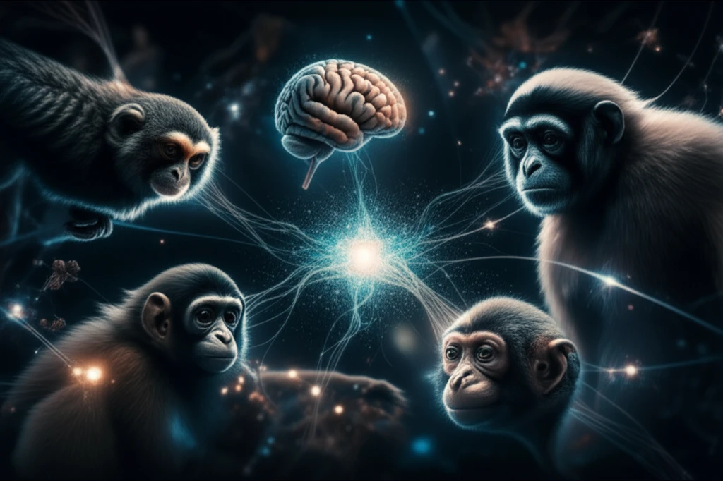
For centuries, scientists have sought to understand the complexities of the human brain by studying our primate relatives. Comparative neuroanatomy, the study of similarities and differences between human and non-human primate brains, offers invaluable insights into the evolution of our own cognitive abilities and neurological functions. However, traditional methods of brain research have often been laborious, expensive, and limited by the availability of specimens.

Now, a groundbreaking initiative from the Japan Monkey Centre (JMC) is poised to revolutionize the field. The JMC has launched a digital repository of primate brain images, providing researchers worldwide with unprecedented access to a wealth of data. This innovative resource promises to accelerate discoveries in neuroscience, conservation, and veterinary medicine.

This new digital archive addresses many challenges such as the lack of available data on non-experimental primates, preservation issues, and limited accessibility. By providing a centralized, high-resolution collection of primate brain images, the JMC is empowering scientists to explore the intricacies of brain structure and function in ways never before possible.

A Digital Treasure Trove: What the JMC Repository Offers

Digital illustration of primate brains interconnected by neural pathways, symbolizing accessible neuroscience data.

The Japan Monkey Centre Primates Brain Imaging Repository is more than just a collection of images; it's a comprehensive resource designed to facilitate cutting-edge research. The repository contains structural MRI and diffusion tensor images (DTI) from 16 brain samples representing 12 different primate species, including:

These images, obtained using a high-resolution 9.4-T MRI scanner, provide detailed anatomical information about each brain. All data is accessible through the JMC collaborative research program's website.

  • Pygmy Marmoset
  • Owl Monkey
  • White-Fronted Capuchin
  • Crab-Eating Macaque
  • Japanese Macaque
  • Bonnet Macaque
  • Toque Macaque
  • Sykes' Monkey
  • Red-Tailed Monkey
  • Schmidt's Guenon
  • De Brazza's Guenon
  • Lar Gibbon
The JMC repository is making significant contributions to comparative neuroscience research by providing the resources to identify neuroanatomical features, optimizing scanning methods, and aiding veterinary neuroradiology. The high-resolution images allow for detailed analysis of brain structures and connectivity, which can be used to compare and contrast different primate species. The repository also provides a valuable resource for developing new methods of scanning and analyzing primate brains. Finally, the repository can be used to train veterinarians in neuroradiology, helping them to diagnose and treat neurological disorders in primates.

A Future of Discovery: The Repository's Expanding Role

The JMC Primates Brain Imaging Repository represents a significant step forward for comparative neuroscience and primate conservation. By providing open access to high-quality brain imaging data, this initiative is empowering researchers worldwide to unlock the secrets of the primate brain and advance our understanding of human cognition and neurological disorders. As the repository continues to grow, it promises to play an increasingly vital role in shaping the future of neuroscience and conservation efforts.

About this Article -

This article was crafted using a human-AI hybrid and collaborative approach. AI assisted our team with initial drafting, research insights, identifying key questions, and image generation. Our human editors guided topic selection, defined the angle, structured the content, ensured factual accuracy and relevance, refined the tone, and conducted thorough editing to deliver helpful, high-quality information.See our About page for more information.

This article is based on research published under:

DOI-LINK: 10.1007/s10329-018-0694-3, Alternate LINK

Title: The Japan Monkey Centre Primates Brain Imaging Repository For Comparative Neuroscience: An Archive Of Digital Records Including Records For Endangered Species

Subject: Animal Science and Zoology

Journal: Primates

Publisher: Springer Science and Business Media LLC

Authors: Tomoko Sakai, Junichi Hata, Hiroki Ohta, Yuta Shintaku, Naoto Kimura, Yuki Ogawa, Kazumi Sogabe, Susumu Mori, Hirotaka James Okano, Yuzuru Hamada, Shinsuke Shibata, Hideyuki Okano, Kenichi Oishi

Published: 2018-10-24

Everything You Need To Know

1

What is comparative neuroanatomy, and why is it important?

Comparative neuroanatomy is the study of the similarities and differences between the brains of different species, particularly human and non-human primates. It's important because it offers insights into the evolution of cognitive abilities and neurological functions. By studying primate brains, scientists can gain a better understanding of how the human brain evolved and how different brain structures contribute to behavior and cognition. While this field focuses on brain structures, future research could explore the genetic and developmental mechanisms that underpin these differences, providing a more complete picture of brain evolution.

2

What types of data are available in the Japan Monkey Centre Primates Brain Imaging Repository?

The Japan Monkey Centre Primates Brain Imaging Repository offers structural MRI and diffusion tensor images (DTI) from 16 brain samples representing 12 different primate species, including Pygmy Marmoset, Owl Monkey, White-Fronted Capuchin, Crab-Eating Macaque, Japanese Macaque, Bonnet Macaque, Toque Macaque, Sykes' Monkey, Red-Tailed Monkey, Schmidt's Guenon, De Brazza's Guenon, and Lar Gibbon. These images provide detailed anatomical information about each brain, captured using a high-resolution 9.4-T MRI scanner. While the repository currently focuses on structural and diffusion imaging, future expansions could include functional MRI (fMRI) data to offer insights into brain activity and connectivity.

3

How does the Japan Monkey Centre Primates Brain Imaging Repository help with primate conservation?

The Japan Monkey Centre Primates Brain Imaging Repository aids primate conservation by providing a centralized, high-resolution collection of primate brain images. This resource addresses the challenges of limited data availability, preservation issues, and accessibility that have historically hampered brain research. By enabling scientists to study primate brains non-invasively, the repository reduces the need for physical specimens, promoting conservation efforts. Furthermore, the repository can be used to train veterinarians in neuroradiology, improving the diagnosis and treatment of neurological disorders in primates, which can contribute to their overall health and well-being. This approach complements field studies that focus on primate behavior and ecology.

4

In what ways can the Japan Monkey Centre Primates Brain Imaging Repository contribute to veterinary medicine?

The Japan Monkey Centre Primates Brain Imaging Repository can contribute significantly to veterinary medicine, particularly in the field of neuroradiology. By providing high-resolution images of primate brains, the repository can be used to train veterinarians in the diagnosis and treatment of neurological disorders in primates. This can lead to improved care for primates in captivity and in the wild. For instance, veterinarians can use the repository's images to identify brain tumors, infections, or injuries in primates, and to develop appropriate treatment plans. The repository also facilitates the development of new methods of scanning and analyzing primate brains, which can further enhance veterinary diagnostic capabilities. This resource augments traditional veterinary practices, offering a more detailed understanding of primate neurological health.

5

What are the potential implications of having open access to high-quality primate brain imaging data through the Japan Monkey Centre?

Open access to high-quality primate brain imaging data through the Japan Monkey Centre has several potential implications. It democratizes neuroscience research by allowing scientists worldwide to explore the intricacies of brain structure and function in ways never before possible, accelerating discoveries related to human cognition and neurological disorders. The repository can also foster collaboration and data sharing among researchers, leading to more comprehensive and robust findings. Furthermore, the availability of this data can promote transparency and reproducibility in neuroscience research, as researchers can independently verify and build upon existing findings. This approach complements traditional research methods by providing a readily accessible and standardized dataset for comparative analyses.

Newsletter Subscribe

Subscribe to get the latest articles and insights directly in your inbox.