Miniature microscope imaging glowing neural networks in the brain

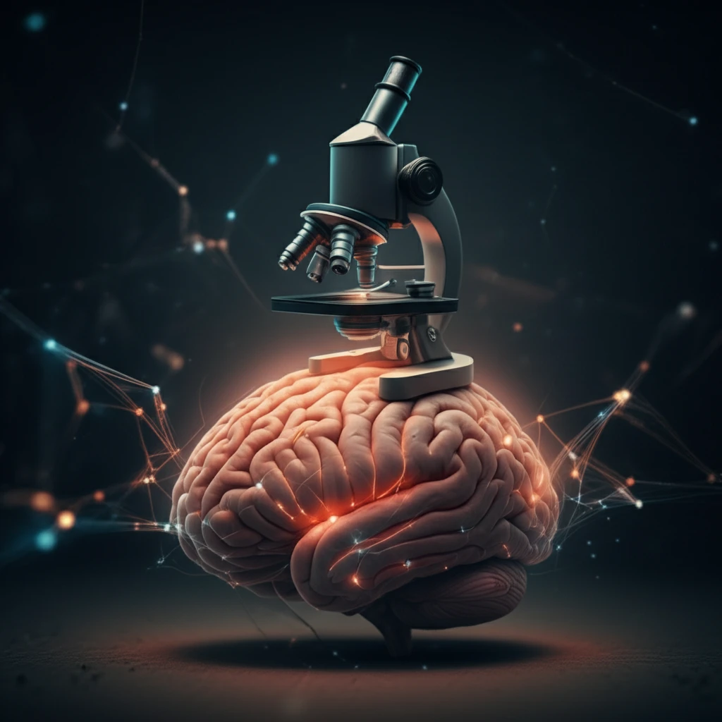
Unlock Precision Brain Imaging: A Breakthrough in Minimally Invasive Techniques

"Discover how new methods in skin suturing and viral infusion enhance neuronal activity imaging with miniature microscopes, improving outcomes in neurological research."


Understanding the brain's complex functions requires tools that can observe neuronal activity on a large scale. Traditional methods like multi-array electrode recordings and two-photon imaging have limitations, either in cell identification or in restricting natural behaviors. Recent advances in miniature one-photon microscopes offer a promising solution, allowing researchers to study brain activity in freely moving animals with cell-type specificity.

However, using these microscopes presents unique challenges. Inserting lenses directly into the brain can cause significant tissue damage, while maintaining clear imaging windows over time is technically demanding. Inflammation and tissue overgrowth can obscure the view, reducing the quality and duration of observable neural activity.

To address these issues, a novel approach combines skin suturing techniques with cortical surface viral infusion to improve both the clarity and longevity of cranial windows. This method minimizes tissue damage, enhances viral labeling precision, and ultimately increases the success rate of imaging neuronal ensembles with head-mounted microscopes.

What Makes This New Brain Imaging Technique Groundbreaking?

Miniature microscope imaging glowing neural networks in the brain

The new technique focuses on two primary innovations: cortical surface viral infusion and scalp skin suturing. Instead of directly injecting viruses into the cortex, a wide-diameter glass pipette is used to gently infuse the viral calcium reporter AAV-GCaMP6 onto the cortical surface. Post-infusion, the scalp skin is sutured over the cranial window to promote recovery and protect against inflammation.

This approach differs significantly from traditional methods that often involve direct intracortical injections and leaving the scalp open. These older techniques can lead to greater tissue damage and a higher risk of complications. The suturing, in particular, provides a protective barrier, reducing inflammation and maintaining a clearer optical window for a longer period.

  • Reduced Tissue Damage: By infusing the virus on the cortical surface, the method avoids deep needle insertions, minimizing damage to brain tissue.
  • Improved Optical Clarity: Suturing the scalp helps maintain a clear imaging window by reducing inflammation and tissue overgrowth.
  • Enhanced Neuronal Labeling: The surface infusion technique preferentially labels superficial cortical layers, which are crucial for integrating neural signals.
  • Increased Success Rate: This combined approach significantly increases the experimental success rate compared to intracortical injections and open-scalp recovery.
The suturing of the scalp ensures that the newly implanted window recovers protected, leading to reduced inflammatory reactions. The large-diameter pipette used in the cortical surface infusion increases the lateral coverage of neuronal labeling, balancing labeling depth and volume to efficiently target neurons in superficial layers, making this technique both efficient and reproducible.

The Future of Brain Imaging

By combining cortical surface viral infusion with post-surgical scalp suturing, researchers can achieve clearer, more stable cranial windows, facilitating long-term studies of neuronal activity. This improved technique promises to advance our understanding of brain functions and offers potential benefits for treating neurological disorders.

About this Article -

This article was crafted using a human-AI hybrid and collaborative approach. AI assisted our team with initial drafting, research insights, identifying key questions, and image generation. Our human editors guided topic selection, defined the angle, structured the content, ensured factual accuracy and relevance, refined the tone, and conducted thorough editing to deliver helpful, high-quality information.See our About page for more information.

Everything You Need To Know

1

What are the primary challenges that current brain imaging techniques face?

Traditional methods, such as multi-array electrode recordings and two-photon imaging, have limitations. These include difficulties in identifying specific cells or restricting the natural behaviors of the subject. Moreover, techniques involving direct lens insertion can cause significant tissue damage, and maintaining clear imaging windows over extended periods is technically challenging. Inflammation and tissue overgrowth often obscure the view, reducing the quality and duration of observable neural activity, hindering long-term studies and precise neuronal activity imaging.

2

How does the new technique improve upon traditional brain imaging methods?

The innovative approach combines cortical surface viral infusion with scalp skin suturing. Instead of direct intracortical injections, the new method uses a wide-diameter glass pipette to infuse the viral calcium reporter AAV-GCaMP6 onto the cortical surface. Post-infusion, the scalp skin is sutured over the cranial window. This minimizes tissue damage, enhances the precision of viral labeling, reduces inflammation, and extends the duration of observable neural activity compared to methods involving direct injections and open-scalp recovery.

3

What is the role of cortical surface viral infusion in this new brain imaging technique?

Cortical surface viral infusion is a key component of the new technique. It involves using a wide-diameter glass pipette to gently infuse the viral calcium reporter AAV-GCaMP6 onto the cortical surface, rather than injecting viruses directly into the cortex. This approach reduces tissue damage because it avoids deep needle insertions. It also enhances neuronal labeling in superficial cortical layers, which are crucial for integrating neural signals. This method improves the overall efficiency and reproducibility of targeting neurons in the superficial layers.

4

Why is skin suturing important in this new brain imaging technique?

Skin suturing plays a vital role by providing a protective barrier over the cranial window. It promotes recovery, reduces inflammation, and prevents tissue overgrowth. The suturing helps maintain a clear optical window, which is essential for high-quality and long-duration neuronal activity imaging. This approach significantly increases the experimental success rate compared to traditional methods, which often leave the scalp open, increasing the risk of complications and reducing the longevity of the imaging window.

5

What are the potential implications of this new brain imaging technique for neurological research and treatment?

By combining cortical surface viral infusion with post-surgical scalp suturing, researchers can achieve clearer and more stable cranial windows. This facilitates long-term studies of neuronal activity, leading to a better understanding of brain functions. This improved technique offers potential benefits for treating neurological disorders by enabling more detailed and prolonged observation of brain activity, which may lead to more targeted and effective treatments. The advancements in the precision of brain imaging have the potential to transform how neurological disorders are studied and treated.

Newsletter Subscribe

Subscribe to get the latest articles and insights directly in your inbox.