Illustration of Affimers and DNA strands interacting within a cell under a super-resolution microscope.

Unlock Cellular Secrets: How DNA-PAINT and Affimers are Revolutionizing Microscopy

"See the unseen: Combining Affimer technology with DNA-PAINT unlocks unprecedented resolution for cellular imaging, offering new insights into cell biology."


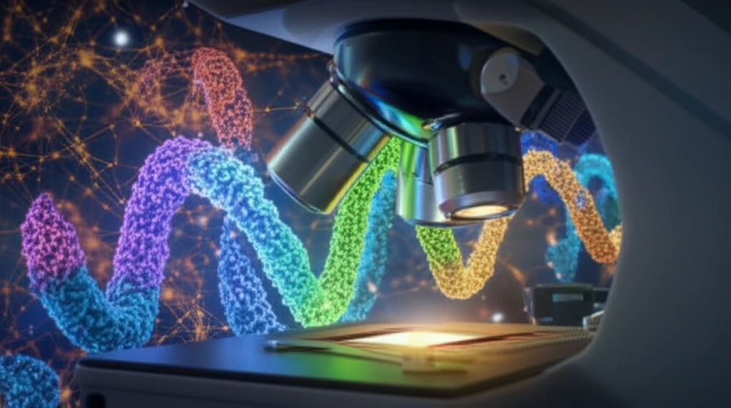
Imagine peering into a cell and seeing its intricate machinery in action, not as a blurry mess, but with crystal clarity. Super-resolution microscopy is making this a reality, pushing past the limitations of traditional light microscopy. We're now able to see details at the nanometer scale, revealing the secrets of cellular processes like never before.

But there's a catch! Getting those super-resolution images isn't always straightforward. The probes we use to tag molecules can be bulky, limiting how closely we can see them. Think of it like trying to assemble a delicate watch with oversized gloves – precision becomes a challenge. Traditional antibodies, while commonly used, can introduce significant distance between the fluorescent marker and the target molecule.

Enter Affimers, a new class of small, highly specific binding agents. Scientists are now combining these Affimers with a technique called DNA-PAINT to achieve even greater resolution in cellular imaging. This innovative approach promises to overcome the limitations of traditional probes, opening up exciting possibilities for biological research and medical diagnostics.

What are Affimers and Why are They a Game Changer?

Illustration of Affimers and DNA strands interacting within a cell under a super-resolution microscope.

Affimers are small proteins engineered to bind to specific target molecules with high affinity. What makes them special? They're much smaller than antibodies, which means they can get closer to the action within a cell. This reduces the 'distance error' – the gap between where the probe binds and where the molecule actually is.

Think of Affimers as tiny, precision-guided missiles compared to the larger, less maneuverable battleships of traditional antibodies. This smaller size also allows for denser labeling, packing more markers into a given area for a brighter, more detailed image. Plus, Affimers can be produced in vitro, meaning they can be developed quickly and efficiently for a wide range of targets.

Here's why Affimers are becoming the darling of super-resolution microscopy:
  • Small Size: Reduced distance error for higher resolution.
  • High Specificity: Precise targeting of desired molecules.
  • Efficient Production: Faster development compared to traditional antibodies.
  • Versatile: Can be engineered to bind to a wide range of targets.
One particularly exciting application is combining Affimers with DNA-PAINT (Points Accumulation for Imaging in Nanoscale Topography). DNA-PAINT is a super-resolution technique that relies on the transient binding of short, fluorescently labeled DNA strands ('imagers') to complementary 'docking' strands attached to the target molecule. The blinking of these imagers allows researchers to pinpoint the location of the target with nanoscale precision.

The Future is Bright: Applications and Implications

The combination of Affimers and DNA-PAINT is poised to revolutionize our understanding of cellular processes. By providing unprecedented detail, this approach can help us to: <ul> <li><b>Understand Disease Mechanisms:</b> Visualize how proteins interact in disease states.</li> <li><b>Develop Targeted Therapies:</b> Identify specific drug targets with greater precision.</li> <li><b>Advance Personalized Medicine:</b> Tailor treatments based on an individual's unique molecular profile.</li> <li><b>Create New Diagnostic Tools:</b> Develop highly sensitive assays for early disease detection.</li> </ul> While challenges remain, such as optimizing labeling strategies and expanding the range of available Affimers, the potential of this technology is undeniable. Get ready for a new era of cellular exploration, where seeing is truly believing!

About this Article -

This article was crafted using a human-AI hybrid and collaborative approach. AI assisted our team with initial drafting, research insights, identifying key questions, and image generation. Our human editors guided topic selection, defined the angle, structured the content, ensured factual accuracy and relevance, refined the tone, and conducted thorough editing to deliver helpful, high-quality information.See our About page for more information.

Everything You Need To Know

1

What are Affimers, and why are they considered a significant advancement in microscopy?

Affimers are small proteins engineered to bind to specific target molecules. Their significance lies in their small size, which allows them to get closer to the target molecule within a cell compared to traditional antibodies. This reduces the 'distance error', leading to higher resolution imaging. Affimers can also be produced efficiently for various targets, making them versatile tools for research and diagnostics. They are important because they reduce the gap between the probe and the target molecule's actual location. This allows for more precise visualization of cellular structures and processes.

2

Can you explain what DNA-PAINT is and how it enables super-resolution microscopy?

DNA-PAINT (Points Accumulation for Imaging in Nanoscale Topography) is a super-resolution microscopy technique. It works by using short, fluorescently labeled DNA strands ('imagers') that transiently bind to complementary 'docking' strands attached to the target molecule. This transient binding causes the imagers to blink, allowing researchers to pinpoint the target's location with nanoscale precision. It is important because it allows for the visualization of cellular structures at a much higher resolution than traditional light microscopy.

3

How does combining Affimers and DNA-PAINT improve cellular imaging, and what are the potential implications?

Combining Affimers with DNA-PAINT allows scientists to visualize cellular structures at the single-molecule level with unprecedented resolution. Affimers provide precise targeting due to their small size and high specificity, while DNA-PAINT enables nanoscale localization of these targets. The implications are significant: this approach allows for a deeper understanding of disease mechanisms, the development of targeted therapies and personalized medicine, and the creation of new diagnostic tools.

4

In what ways can Affimers and DNA-PAINT contribute to a better understanding of disease mechanisms?

The combination of Affimers and DNA-PAINT can help us understand how proteins interact in disease states. Visualizing these interactions at high resolution can reveal the underlying causes of diseases and identify potential drug targets. For example, scientists can observe how misfolded proteins aggregate in neurodegenerative diseases or how cancer cells evade the immune system. This information is crucial for developing effective treatments.

5

Are there any limitations to using Affimers and DNA-PAINT in cellular imaging?

Yes, the use of Affimers and DNA-PAINT does have some limitations. Optimizing labeling strategies can be challenging, as it requires careful selection of Affimers and DNA sequences to ensure efficient and specific binding. Additionally, expanding the range of available Affimers to target a wider variety of molecules is an ongoing effort. Despite these challenges, the potential of this technology for advancing biological research and medical diagnostics is substantial.

Newsletter Subscribe

Subscribe to get the latest articles and insights directly in your inbox.