Surreal illustration of a protein illuminating a cancer tumor.

Unlock Cancer's Secrets: How New Imaging Tech Could Revolutionize Treatment

"A groundbreaking protein promises faster, more accurate detection of cancer's hidden vulnerabilities."


Cancer's ability to evade the immune system is a major obstacle in treatment. Immune checkpoint inhibitors, which unleash the body's natural defenses, have shown promise, but predicting which patients will respond remains a challenge. Traditional methods like biopsies have limitations, leading researchers to explore innovative imaging techniques.

Positron Emission Tomography (PET) imaging offers a non-invasive way to visualize cancer at the molecular level. However, current antibody-based PET tracers have drawbacks, including slow clearance from the body and restrictions on radioisotope use. This has spurred the development of smaller protein tracers that can provide faster, clearer images.

Now, a team of scientists has engineered a novel small protein for PET imaging that targets Programmed Death Ligand-1 (PD-L1), a key player in cancer's immune evasion. This innovative tracer, validated in mouse models and human cancer tissues, holds the potential to revolutionize cancer diagnosis and treatment strategies.

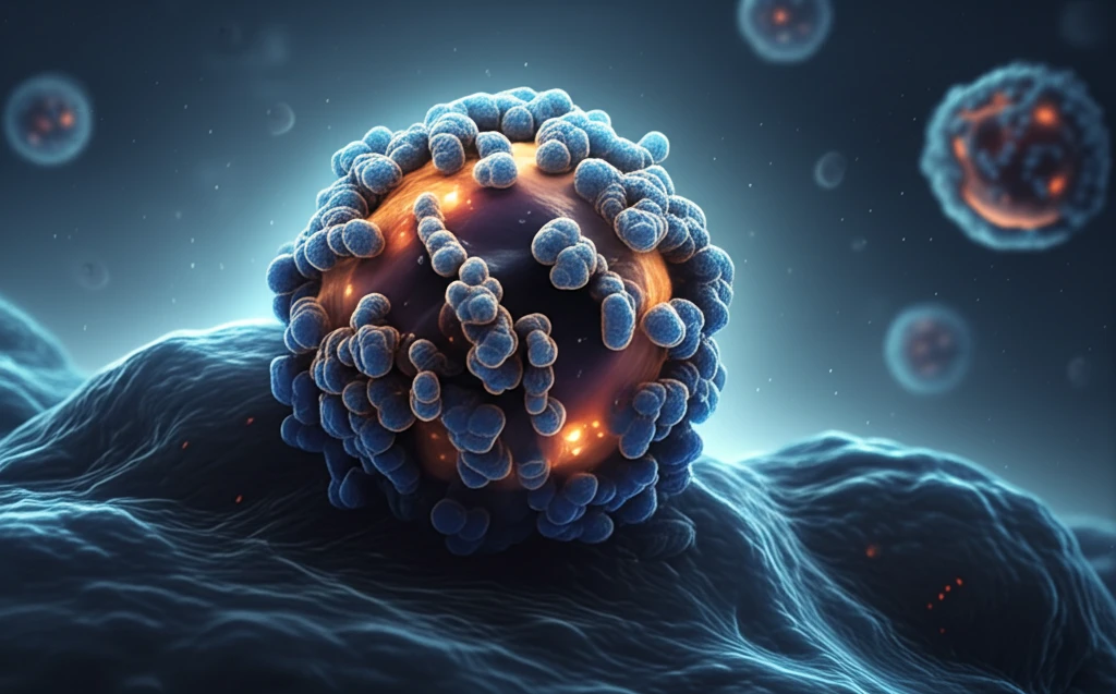
What is PD-L1 and Why is it Important for Cancer Treatment?

Surreal illustration of a protein illuminating a cancer tumor.

PD-L1, or Programmed Death-Ligand 1, is a protein found on the surface of cells, including cancer cells. It interacts with PD-1, another protein found on immune cells called T-cells. This interaction acts as a 'brake' on the immune system, preventing T-cells from attacking and destroying cancer cells. In essence, PD-L1 helps cancer cells hide from the immune system.

High levels of PD-L1 are often found in aggressive cancers, allowing them to effectively evade immune surveillance. This makes PD-L1 a crucial target for cancer immunotherapy. By blocking the interaction between PD-L1 and PD-1, immune checkpoint inhibitors can release the brakes on the immune system, enabling T-cells to recognize and kill cancer cells.

  • Understanding PD-L1 levels helps predict responses to immunotherapy.
  • PD-L1 expression can vary within a tumor and change over time.
  • Accurate PD-L1 detection is essential for personalized treatment strategies.
However, current methods for assessing PD-L1 expression, primarily immunohistochemistry (IHC) on biopsy samples, have limitations. Biopsies only capture a small snapshot of the tumor and may not represent the entire picture. This is where new imaging techniques like PET imaging with the engineered protein tracer come into play.

The Future of Cancer Diagnosis: Personalized and Precise

The development of this novel PET tracer represents a significant step forward in cancer imaging. By providing a non-invasive, accurate, and dynamic assessment of PD-L1 expression, this technology has the potential to transform cancer diagnosis and treatment decisions. It could help identify patients most likely to benefit from immunotherapy, optimize treatment strategies, and ultimately improve outcomes for those battling cancer.

About this Article -

This article was crafted using a human-AI hybrid and collaborative approach. AI assisted our team with initial drafting, research insights, identifying key questions, and image generation. Our human editors guided topic selection, defined the angle, structured the content, ensured factual accuracy and relevance, refined the tone, and conducted thorough editing to deliver helpful, high-quality information.See our About page for more information.

Everything You Need To Know

1

What is Programmed Death Ligand-1 (PD-L1), and why is it a significant target in cancer treatment?

Programmed Death Ligand-1, or PD-L1, is a protein present on the surface of cells, including cancer cells, that interacts with Programmed Death-1 (PD-1) on T-cells. This interaction acts like a 'brake' on the immune system, preventing T-cells from attacking cancer cells. PD-L1 helps cancer cells evade immune detection, making it a crucial target. High PD-L1 levels are found in aggressive cancers. Blocking the PD-L1 and PD-1 interaction with immune checkpoint inhibitors releases this brake, enabling T-cells to recognize and kill cancer cells. Assessing PD-L1 levels is crucial for predicting responses to immunotherapy and tailoring personalized treatment strategies. Current methods like immunohistochemistry (IHC) on biopsies have limitations, prompting the exploration of advanced imaging techniques.

2

How does the new protein tracer used in Positron Emission Tomography (PET) imaging improve the detection of cancer vulnerabilities compared to traditional methods like biopsies?

The novel small protein engineered for PET imaging offers a non-invasive method to visualize Programmed Death Ligand-1 (PD-L1) expression at the molecular level, addressing the limitations of traditional biopsies. Unlike biopsies, which only capture a small snapshot of the tumor and may not represent the entire picture, PET imaging with this tracer provides a comprehensive and dynamic assessment of PD-L1 expression throughout the tumor. This allows for a more accurate determination of whether a patient is likely to respond to immunotherapy. Current antibody-based PET tracers have drawbacks, including slow clearance from the body and restrictions on radioisotope use. The innovative tracer, validated in mouse models and human cancer tissues, holds the potential to revolutionize cancer diagnosis and treatment strategies.

3

What are immune checkpoint inhibitors, and how does Programmed Death Ligand-1 (PD-L1) influence their effectiveness in cancer treatment?

Immune checkpoint inhibitors are drugs that unleash the body's natural defenses to fight cancer. They work by blocking specific proteins, such as Programmed Death-1 (PD-1) or Programmed Death Ligand-1 (PD-L1), that help cancer cells evade the immune system. PD-L1, found on cancer cells, binds to PD-1 on T-cells, inhibiting the T-cells' ability to attack the cancer. When immune checkpoint inhibitors block this interaction, they release the 'brake' on the immune system, allowing T-cells to recognize and destroy cancer cells. The level of PD-L1 expression in a tumor can influence how well these inhibitors work; higher PD-L1 levels often indicate a greater potential response to treatment, making accurate PD-L1 detection crucial for personalized treatment strategies.

4

What are the potential benefits of using this new protein for Programmed Death Ligand-1 (PD-L1) PET imaging in developing personalized cancer therapies?

The new protein for Programmed Death Ligand-1 (PD-L1) PET imaging offers several key benefits in developing personalized cancer therapies. It allows for a non-invasive, accurate, and dynamic assessment of PD-L1 expression, helping identify patients most likely to benefit from immunotherapy. It enables real-time monitoring of treatment response, facilitating timely adjustments to therapy. The enhanced accuracy in PD-L1 detection supports more informed treatment decisions, optimizing strategies for individual patients. This leads to improved outcomes for those battling cancer. The new imaging agent is designed for faster and clearer images. By providing a comprehensive view of PD-L1 expression, this technology helps tailor treatments to the unique characteristics of each patient's cancer, moving towards more effective and personalized cancer care.

5

How might the ability to dynamically assess Programmed Death Ligand-1 (PD-L1) expression using new PET imaging techniques impact cancer treatment monitoring and adjustment?

The dynamic assessment of Programmed Death Ligand-1 (PD-L1) expression through new PET imaging techniques has significant implications for cancer treatment monitoring and adjustment. Traditional methods like biopsies provide only a static snapshot of PD-L1 levels, which can change over time and may not represent the entire tumor. PET imaging, on the other hand, offers a non-invasive way to repeatedly assess PD-L1 expression during treatment. This allows clinicians to monitor how PD-L1 levels respond to therapy, helping them determine if the treatment is effective or if adjustments are needed. If PD-L1 expression decreases, it may indicate a positive response, while stable or increasing levels might suggest the need for alternative strategies. This dynamic monitoring enables more informed and timely treatment decisions, potentially improving patient outcomes. This PET imaging allows repeated tests unlike traditional testing methods.

Newsletter Subscribe

Subscribe to get the latest articles and insights directly in your inbox.