Illustration of a preduodenal portal vein compressing the duodenum in an infant with heterotaxy syndrome.

Unforeseen Obstruction: How a Rare Condition Complicated Infant Duodenal Surgery

"Discover how surgeons navigated a heterotaxy syndrome case with a preduodenal portal vein, causing unexpected pulmonary venous obstruction during a routine procedure."


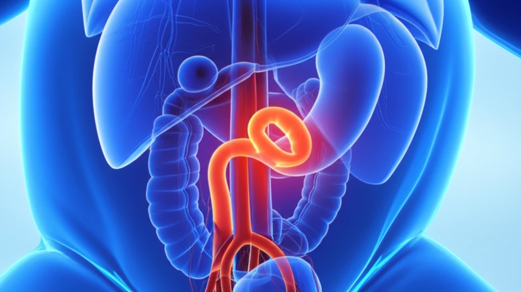
Heterotaxy syndrome (HS) is a rare congenital condition characterized by a broad spectrum of anatomical abnormalities, particularly affecting the heart and abdominal organs. These abnormalities can include pulmonary valve stenosis, interruption of the inferior vena cava, total anomalous pulmonary venous connection (TAPVC), and malformations such as asplenia and polysplenia. Intestinal malrotation and the presence of a preduodenal portal vein (PDPV) are also frequently observed in individuals with HS.

In cases where infracardiac TAPVC is present, a complex of pulmonary veins converges and drains into a vertical vein, which then connects to the systemic venous system below the diaphragm. The most common drainage site for this vertical vein is the portal vein. PDPV, another rare anomaly, occurs when the portal vein passes anterior to the duodenum instead of its usual posterior position. This condition is strongly associated with heterotaxy and situs inversus and may lead to duodenal obstruction.

A recent case highlights the complexities involved in managing HS. An infant with infracardiac TAPVC and PDPV experienced repeated episodes of hemodynamic instability during urgent surgery for duodenal obstruction. The surgical team's ability to identify and address the unexpected source of these complications underscores the importance of vigilance and comprehensive understanding of rare anatomical variations in HS patients.

The Case: Unexpected Twists During Duodenal Surgery

Illustration of a preduodenal portal vein compressing the duodenum in an infant with heterotaxy syndrome.

A 3-day-old boy, prenatally diagnosed with heterotaxy syndrome (HS), was scheduled for urgent laparotomy to address duodenal atresia. His condition included an interrupted inferior vena cava, a single right ventricle, pulmonary valve stenosis, and infracardiac TAPVC. Initial assessments suggested a relatively mild preoperative pulmonary venous obstruction (PVO), leading the team to proceed with the surgery.

The laparotomy revealed intestinal malrotation, characterized by Ladd's bands. As the surgeons worked to correct the duodenal obstruction, the infant experienced repetitive episodes of severe hypotension and hypoxia. Standard interventions proved ineffective, prompting a thorough re-evaluation of the surgical field.

  • Initial Presentation: 3-day-old infant with HS, duodenal atresia, and infracardiac TAPVC.
  • Surgical Finding: Intestinal malrotation with Ladd's bands.
  • Complications: Repetitive severe hypotension and hypoxia during surgery.
  • Key Discovery: A greatly dilated PDPV compressing the duodenum.
Further examination revealed a greatly dilated PDPV crossing over and compressing the proximal duodenum externally. The surgical team hypothesized that surgical manipulation was directly compressing the dilated PDPV, into which the TAPVC drained, leading to pulmonary venous obstruction (PVO). This was confirmed by a postoperative computed tomography scan showing that the vertical vein from the pulmonary venous confluence drained into the portal vein. A Ladd procedure with gastroduodenostomy was performed, bypassing the PDPV. Postoperatively, the infant was admitted to intensive care and stabilized with inotropic support. Subsequent imaging confirmed the vertical vein drained into the portal vein.

Clinical Implications and Recommendations

The case highlights the critical importance of considering PDPV in HS patients undergoing surgery for intestinal malrotation and duodenal obstruction, especially those with infracardiac TAPVC. Both pediatric surgeons and anesthesiologists must be aware of the potential for pulmonary venous drainage into the PDPV. Anticipating and recognizing this rare anatomical variation can help prevent catastrophic PVO during surgery.

About this Article -

This article was crafted using a human-AI hybrid and collaborative approach. AI assisted our team with initial drafting, research insights, identifying key questions, and image generation. Our human editors guided topic selection, defined the angle, structured the content, ensured factual accuracy and relevance, refined the tone, and conducted thorough editing to deliver helpful, high-quality information.See our About page for more information.

Everything You Need To Know

1

What is Heterotaxy Syndrome (HS) and what are its common associated anomalies?

Heterotaxy Syndrome (HS) is a rare congenital condition that causes a wide range of anatomical abnormalities. These anomalies often affect the heart and abdominal organs. Commonly associated conditions include pulmonary valve stenosis, interruption of the inferior vena cava, and total anomalous pulmonary venous connection (TAPVC). Furthermore, malformations such as asplenia and polysplenia can occur. Intestinal malrotation and the presence of a preduodenal portal vein (PDPV) are also frequently seen in individuals with HS.

2

What role did the Preduodenal Portal Vein (PDPV) play in the complications during the infant's duodenal surgery?

In this case, the Preduodenal Portal Vein (PDPV) significantly complicated the surgery. The infant had a dilated PDPV that was compressing the proximal duodenum externally. The surgical team hypothesized that manipulation during the Ladd procedure was compressing the PDPV. As the TAPVC drained into the portal vein, this compression led to pulmonary venous obstruction (PVO), causing repetitive episodes of severe hypotension and hypoxia. The PDPV's unusual position, crossing anterior to the duodenum, directly caused the unexpected obstruction and hemodynamic instability.

3

How does Total Anomalous Pulmonary Venous Connection (TAPVC) relate to the complications in this specific heterotaxy syndrome case?

In this case, the infant had infracardiac TAPVC. Infracardiac TAPVC involves a complex arrangement where the pulmonary veins converge and drain into a vertical vein. This vertical vein connects to the systemic venous system below the diaphragm, often draining into the portal vein. The presence of TAPVC and the subsequent drainage into the Preduodenal Portal Vein (PDPV) meant that any compression of the PDPV could obstruct pulmonary venous flow, leading to pulmonary venous obstruction (PVO), which was the underlying cause of the hemodynamic instability observed during surgery. The unusual venous drainage pathway exacerbated the effects of the PDPV.

4

Why is it crucial for surgeons and anesthesiologists to be aware of Preduodenal Portal Vein (PDPV) in patients with Heterotaxy Syndrome (HS) and Total Anomalous Pulmonary Venous Connection (TAPVC)?

Awareness of the Preduodenal Portal Vein (PDPV) is critical for pediatric surgeons and anesthesiologists, especially when treating Heterotaxy Syndrome (HS) patients with Total Anomalous Pulmonary Venous Connection (TAPVC) because this anatomical variation poses a significant risk during surgery for intestinal malrotation and duodenal obstruction. If the PDPV is not considered, surgical manipulations can inadvertently compress the vein, leading to pulmonary venous obstruction (PVO). This obstruction can cause severe hypotension and hypoxia, potentially leading to life-threatening complications. Recognizing this rare anomaly allows the surgical team to anticipate and mitigate potential complications, improving patient outcomes by adjusting surgical techniques to avoid compression and ensuring appropriate monitoring and management.

5

What specific surgical interventions were performed, and how did they address the complications caused by the Preduodenal Portal Vein (PDPV)?

The primary surgical intervention performed was a Ladd procedure with gastroduodenostomy. This approach was chosen to correct the duodenal obstruction caused by the intestinal malrotation and bypass the Preduodenal Portal Vein (PDPV). The team recognized that compression of the dilated PDPV, where the pulmonary venous confluence drained due to the infracardiac TAPVC, was causing the hemodynamic instability. By bypassing the PDPV, the surgical team aimed to alleviate the external compression on the vein and improve pulmonary venous flow, thereby resolving the pulmonary venous obstruction (PVO) and stabilizing the infant's condition. Postoperative imaging confirmed the success of redirecting venous flow away from the compression site.

Newsletter Subscribe

Subscribe to get the latest articles and insights directly in your inbox.