Uncommon Anatomy, Unexpected Danger: When Your Kidneys Aren't Where They Should Be
"A Case Study Reveals How Bilateral Renal Malrotation Can Lead to Acute Pyelonephritis and Septic Shock, Highlighting the Need for Vigilant Diagnosis."
Imagine your body's organs are like puzzle pieces, each fitting perfectly to ensure smooth functioning. Now, picture one of those pieces—a kidney—slightly out of place. This isn't just a minor anatomical quirk; it can lead to serious health problems. Bilateral renal malrotation, a rare condition where both kidneys are abnormally rotated, can create a perfect storm for infections and other complications.
A recent case study published in the Chonnam Medical Journal sheds light on this often-overlooked condition. The study details the experience of a 71-year-old woman who presented with seemingly straightforward symptoms: left flank pain, oliguria (decreased urine output), and hypotension (low blood pressure). Initial assessments pointed towards a common culprit: pyelonephritis, a kidney infection. However, the underlying cause turned out to be far more complex.
This case underscores the critical importance of considering rare anatomical variations when diagnosing and treating seemingly common conditions. It also highlights the power of advanced imaging techniques in uncovering hidden health risks. As we delve into this case, you'll gain a deeper understanding of renal malrotation, its potential dangers, and the diagnostic steps that can save lives.
The Case Unfolds: Symptoms, Diagnosis, and the Role of Imaging
The 71-year-old patient's initial symptoms were concerning but not particularly unique. Flank pain is a common complaint, and the presence of oliguria and hypotension suggested a systemic issue. Pyuria (pus in the urine) and severe sepsis quickly led to a diagnosis of acute pyelonephritis, a bacterial infection of the kidneys. Standard treatment protocols were initiated, but the underlying cause remained a mystery.
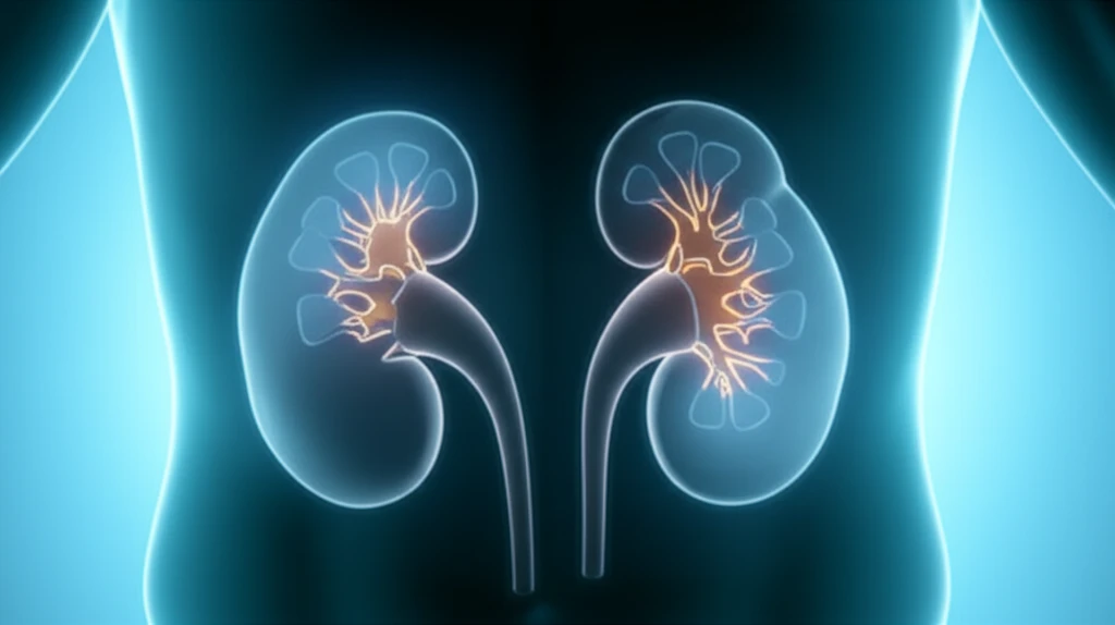
- CT Scan Insights: The CT scan also revealed that the right kidney was positioned 5 cm lower than the left kidney, indicating renal ectopia (abnormal location). Furthermore, mild dilation was observed in the proximal ureter of the left kidney, suggesting a potential obstruction.
- 3D Reconstruction: To gain a clearer understanding of the kidney's spatial arrangement, a three-dimensional computed tomography reconstruction image was created. This image vividly displayed the anteriorly rotated kidneys, confirming the extent of the malrotation.
The Takeaway: Vigilance and Advanced Imaging for Rare Conditions
This case serves as a powerful reminder that even common conditions can have uncommon underlying causes. Bilateral renal malrotation, though rare, can significantly increase the risk of pyelonephritis and subsequent septic shock. The study underscores the importance of considering anatomical variations in patients with recurrent UTIs or atypical kidney-related symptoms. Advanced imaging techniques, such as CT intravenous pyelograms and 3D reconstructions, play a vital role in identifying these variations and guiding appropriate treatment strategies. For patients and healthcare providers alike, this case highlights the need for vigilance, thorough investigation, and a willingness to consider the less obvious possibilities when faced with complex medical presentations.