Surreal illustration of a leg transitioning into an MRI scan for DVT visualization.

Unclogging the Mystery: Can Advanced Imaging Solve Deep Vein Thrombosis?

"New research explores how Magnetic Resonance Imaging (MRI) could revolutionize the diagnosis and treatment of deep vein thrombosis, offering hope for personalized care and fewer complications."


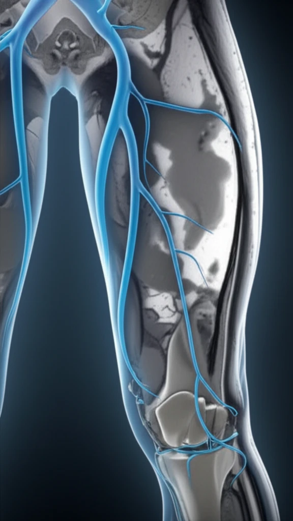
Deep vein thrombosis (DVT), a condition where blood clots form in the deep veins of the body (usually the legs), is a serious health concern. If left untreated, DVT can lead to pulmonary embolism, a life-threatening condition where the clot travels to the lungs. Current diagnostic methods often rely on factors like patient history and physical exams, which aren't always accurate.

Traditional treatments such as catheter-directed thrombolysis (CDT), which involves using a catheter to deliver clot-dissolving drugs, can be effective, but also carry risks such as bleeding complications. This has created a need for more precise ways to assess the characteristics of blood clots and predict how well they will respond to treatment.

Now, a new study presented at the 19th Meeting of the European Venous Forum explores the use of advanced Magnetic Resonance Imaging (MRI) techniques to assess thrombus composition and predict its 'lysability' or likelihood of dissolving with treatment. This research offers the potential for a more personalized approach to DVT care, helping doctors to choose the right treatment for the right patient.

MSTI: A New Vision for Thrombus Characterization

Surreal illustration of a leg transitioning into an MRI scan for DVT visualization.

The study, led by Justinas Silickas and colleagues at King's College London, focused on a novel MRI technique called multi-sequence thrombus imaging (MSTI). MSTI involves using different MRI sequences to map the thrombus, looking at factors like T1 relaxation times, magnetization transfer, and diffusion-weighted imaging. These parameters provide insights into the age, structure, and composition of the clot.

Researchers enrolled 39 patients with acute iliofemoral DVTs (clots in the iliac and femoral veins). Before thrombolysis, patients underwent MSTI using a 3-Tesla MR scanner. The MSTI scan took around 35 minutes per patient. After imaging, the success of thrombolysis was evaluated using contrast venography, which looked for the absence of residual thrombus and the lack of need for stenting.

  • T1 Relaxation Times: Non-lysed thrombi showed significantly higher T1 relaxation times compared to lysed thrombi (967 ± 20 ms vs. 790 ± 29 ms, P<0.001). This suggests that older, more organized clots have different water content and molecular interactions than fresh clots.
  • Magnetization Transfer (MTR): MTR values were similar in both lysed and non-lysed thrombi, indicating that this parameter alone may not be a good predictor of lysability.
  • Mean ADC: Non-lysed thrombi had higher mean ADC values than lysed thrombi (1.7 ± 0.1 mm²/s vs. 1.4 ± 0.1 mm²/s, P = 0.05). ADC reflects the water diffusion within the thrombus, which can be influenced by its structure and cellular components.
Receiver-operating characteristic (ROC) analysis revealed that T1 mapping had the highest area under the curve (AUC) of 0.81, indicating strong diagnostic accuracy. ADC also showed significant predictive value with an AUC of 0.66. By combining T1, MTR, and ADC values, the researchers achieved a sensitivity of 89% and a specificity of 100% in distinguishing lysed and non-lysed thrombi.

A Promising Step Toward Personalized DVT Treatment

This research highlights the potential of MSTI to improve the management of acute iliofemoral DVTs. By identifying patients with thrombi that are most likely to respond to thrombolysis, doctors can avoid unnecessary treatments and reduce the risk of complications. However, further studies are needed to optimize image analysis methods, validate these findings in other centers, and ultimately improve patient outcomes.

About this Article -

This article was crafted using a human-AI hybrid and collaborative approach. AI assisted our team with initial drafting, research insights, identifying key questions, and image generation. Our human editors guided topic selection, defined the angle, structured the content, ensured factual accuracy and relevance, refined the tone, and conducted thorough editing to deliver helpful, high-quality information.See our About page for more information.

Everything You Need To Know

1

What is Deep Vein Thrombosis (DVT) and why is it a serious health concern?

Deep Vein Thrombosis (DVT) is a condition where blood clots form in the deep veins of the body, most commonly in the legs. It poses a significant health risk because these clots can potentially break away and travel to the lungs, causing a life-threatening condition known as pulmonary embolism. Traditional diagnostic methods and treatments carry risks, underscoring the importance of accurate and personalized treatment strategies.

2

How does Magnetic Resonance Imaging (MRI) fit into the diagnosis and treatment of DVT?

MRI is being explored as a novel diagnostic tool for DVT, specifically using a technique called multi-sequence thrombus imaging (MSTI). MSTI uses various MRI sequences to assess the composition and predict the 'lysability' of a blood clot. This means evaluating how likely the clot is to dissolve with treatment. The goal is to enable doctors to choose the most effective and personalized treatment approach for each patient, reducing the risk of complications.

3

What is MSTI, and how does it help in evaluating blood clots?

MSTI, or multi-sequence thrombus imaging, is a specialized MRI technique that uses different MRI sequences to analyze the characteristics of a blood clot. These sequences measure parameters such as T1 relaxation times, magnetization transfer (MTR), and mean Apparent Diffusion Coefficient (ADC). These parameters offer insights into the age, structure, and composition of the clot, helping to predict how it will respond to treatment. For example, in the study, higher T1 relaxation times correlated with non-lysed thrombi, indicating older, more organized clots.

4

What role did T1 relaxation times, MTR, and ADC play in the study's findings?

In the study, T1 relaxation times, MTR, and ADC were used to characterize the blood clots. Researchers found that T1 mapping showed strong diagnostic accuracy, with higher values indicating clots less likely to dissolve. MTR values were similar in both lysed and non-lyed thrombi, indicating that this parameter may not be a good predictor of lysability. ADC also showed significant predictive value. By combining these values, the study achieved a high sensitivity and specificity in distinguishing between clots that would dissolve and those that would not, leading to a more personalized approach for DVT treatment.

5

What are the potential benefits of using MSTI for DVT treatment, and what are the next steps?

The primary benefit of MSTI is the potential for personalized DVT treatment. By using MSTI, doctors can identify which patients are most likely to respond to thrombolysis, avoiding unnecessary procedures and reducing the risk of complications associated with treatments like catheter-directed thrombolysis (CDT). The next steps involve optimizing image analysis methods, validating these findings in other clinical settings, and conducting further studies to ultimately improve patient outcomes and refine the use of MSTI in DVT management.

Newsletter Subscribe

Subscribe to get the latest articles and insights directly in your inbox.