Surreal illustration of a brain scan visualizing inflammation with glowing immune cells.

Ultra-High Field MRI: A New Window into Inflammation?

"Exploring the potential of 21.1 T MRI for enhanced detection and detail in neuroinflammation studies."


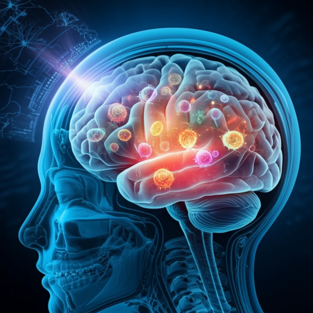
Magnetic Resonance Imaging (MRI) has revolutionized medical diagnostics, but scientists are always looking for ways to improve its sensitivity and resolution. One promising avenue is increasing the strength of the magnetic field used in MRI scanners. This article explores the exciting potential of ultra-high field MRI, specifically using a powerful 21.1 Tesla (T) scanner, to enhance the detection and understanding of inflammatory processes, particularly in the brain.

Traditional MRI methods sometimes struggle to visualize subtle inflammatory changes. Researchers are particularly interested in fluorine-19 (¹⁹F) MRI, which offers unique advantages for tracking cells involved in inflammation. However, the low concentration of fluorine in the body makes it challenging to get clear signals. This is where the increased power of ultra-high field MRI comes into play.

This article delves into a recent study that investigated the benefits of performing ¹⁹F MRI at 21.1 T, compared to a more standard 9.4 T. The focus was on visualizing neuroinflammation in an experimental model, revealing how the enhanced sensitivity of the stronger magnetic field could unlock new insights into inflammatory diseases.

Unveiling the Power of 21.1 T MRI: What the Research Shows

Surreal illustration of a brain scan visualizing inflammation with glowing immune cells.

The research team conducted experiments using perfluoro-15-crown-5-ether (PFCE), a fluorine-containing compound, to study inflammation in mice with experimental autoimmune encephalomyelitis (EAE), a model for multiple sclerosis. They compared MRI scans obtained at both 9.4 T and 21.1 T to assess the gains in signal-to-noise ratio (SNR) and image quality.

Here's a breakdown of the key findings:

  • Enhanced SNR: The 21.1 T MRI showed a significant increase in SNR compared to 9.4 T, both for pure PFCE and PFCE nanoparticles. This means clearer, more detailed images.
  • Improved Resolution: High-resolution ¹⁹F MRI at 21.1 T revealed signals in the brains and lymph nodes of EAE mice that were not detectable at 9.4 T, highlighting the ability to visualize finer details of inflammation.
  • Faster Relaxation: The study also found that the T1 relaxation time of PFCE was shorter at 21.1 T, which could allow for faster scanning and improved image acquisition.
These results suggest that ultra-high field MRI offers a significant advantage for studying inflammatory processes. The enhanced SNR and resolution can help researchers visualize subtle changes, track immune cell migration, and gain a deeper understanding of disease mechanisms.

The Future of Inflammation Imaging: Implications and Possibilities

This research provides compelling evidence for the potential of ultra-high field MRI to revolutionize how we study and diagnose inflammatory diseases. The ability to visualize subtle changes and track immune cell activity with greater precision could lead to earlier diagnosis, more targeted treatments, and a better understanding of disease progression.

While 21.1 T MRI scanners are not yet widely available, this study highlights the value of investing in this technology. As ultra-high field MRI becomes more accessible, we can expect to see significant advances in our understanding and management of a wide range of inflammatory conditions, including neurological disorders, autoimmune diseases, and cancer.

Further research is needed to optimize ¹⁹F MRI techniques at ultra-high field strengths and explore the full potential of this technology for clinical applications. However, the initial findings are promising and suggest that ultra-high field MRI could play a crucial role in the future of inflammation imaging.

About this Article -

This article was crafted using a human-AI hybrid and collaborative approach. AI assisted our team with initial drafting, research insights, identifying key questions, and image generation. Our human editors guided topic selection, defined the angle, structured the content, ensured factual accuracy and relevance, refined the tone, and conducted thorough editing to deliver helpful, high-quality information.See our About page for more information.

This article is based on research published under:

DOI-LINK: 10.1007/s10334-018-0710-z, Alternate LINK

Title: Fluorine-19 Mri At 21.1 T: Enhanced Spin–Lattice Relaxation Of Perfluoro-15-Crown-5-Ether And Sensitivity As Demonstrated In Ex Vivo Murine Neuroinflammation

Subject: Radiology, Nuclear Medicine and imaging

Journal: Magnetic Resonance Materials in Physics, Biology and Medicine

Publisher: Springer Science and Business Media LLC

Authors: Sonia Waiczies, Jens T. Rosenberg, Andre Kuehne, Ludger Starke, Paula Ramos Delgado, Jason M. Millward, Christian Prinz, Joao Dos Santos Periquito, Andreas Pohlmann, Helmar Waiczies, Thoralf Niendorf

Published: 2018-11-12

Everything You Need To Know

1

What is ultra-high field MRI and how does it improve imaging?

Ultra-high field MRI uses stronger magnetic fields, like the 21.1 Tesla (T) scanner, to get better images in Magnetic Resonance Imaging. This higher field strength improves the signal-to-noise ratio (SNR) and resolution, which helps in seeing finer details. It's especially useful for fluorine-19 (¹⁹F) MRI, where the signal is usually weak.

2

What are the key advantages observed when using a 21.1 T MRI compared to a 9.4 T MRI?

The 21.1 T MRI showed a significant increase in signal-to-noise ratio (SNR) compared to the 9.4 T. High-resolution ¹⁹F MRI at 21.1 T revealed signals in the brains and lymph nodes of EAE mice that were not detectable at 9.4 T, highlighting the ability to visualize finer details of inflammation. The T1 relaxation time of PFCE was shorter at 21.1 T, which could allow for faster scanning and improved image acquisition.

3

How does ultra-high field MRI enhance the study of inflammatory processes?

The improved signal-to-noise ratio (SNR) and resolution of ultra-high field MRI, like the 21.1 T, help researchers see subtle inflammatory changes that might be missed by standard methods. Using perfluoro-15-crown-5-ether (PFCE) in experimental autoimmune encephalomyelitis (EAE) studies shows how specific compounds can be tracked to understand disease mechanisms better.

4

Why is fluorine-19 MRI important for inflammation research, and how does ultra-high field MRI help?

Fluorine-19 (¹⁹F) MRI has great potential for tracking cells involved in inflammation due to its unique properties. However, the low concentration of fluorine in the body makes it difficult to get clear signals with traditional MRI. Ultra-high field MRI, using a 21.1 Tesla (T) scanner, addresses this by boosting the signal and improving the visibility of fluorine-containing compounds like perfluoro-15-crown-5-ether (PFCE). This allows researchers to track immune cell migration more effectively.

5

What are the potential implications of ultra-high field MRI for diagnosing and treating inflammatory diseases?

The findings suggest that ultra-high field MRI could lead to earlier and more accurate diagnoses of inflammatory diseases. By visualizing subtle changes and tracking immune cell activity with greater precision, clinicians may be able to detect diseases at earlier stages and monitor their progression more effectively. This could pave the way for more targeted and personalized treatment strategies, ultimately improving patient outcomes.

Newsletter Subscribe

Subscribe to get the latest articles and insights directly in your inbox.