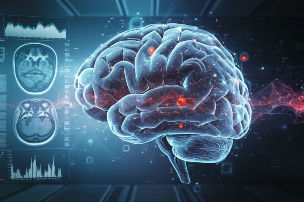
Traumatic Brain Injury: How Cutting-Edge Imaging Techniques Are Revolutionizing Diagnosis and Treatment
"Unveiling the Power of Nuclear Medicine Neuroimaging in Understanding and Treating Brain Trauma"
Traumatic brain injury (TBI) is a major global health concern, affecting millions of people worldwide each year. From sports-related concussions to severe injuries from accidents or military conflicts, TBI can have devastating and long-lasting effects on individuals and their families. Understanding the complexities of TBI and developing effective diagnostic and treatment strategies are critical to improving outcomes for those affected.
Traditional methods of assessing TBI, such as CT scans and MRIs, primarily focus on identifying structural damage to the brain. However, these techniques often fall short in revealing the full extent of the injury, particularly in cases of diffuse axonal injury (DAI) where damage occurs at a microscopic level. This is where nuclear medicine neuroimaging steps in, offering a unique perspective on the functional and molecular changes that occur in the brain after TBI.
This article delves into the world of nuclear medicine neuroimaging and its transformative role in the diagnosis and management of TBI. We'll explore techniques like Positron Emission Tomography (PET) and Single-Photon Emission Computed Tomography (SPECT), highlighting how they provide valuable insights into the metabolic and cellular processes affected by TBI. By understanding these advanced imaging methods, we can better appreciate their potential to revolutionize the way we approach TBI care.
Decoding TBI: How PET Scans Reveal Metabolic Changes in the Brain

Positron Emission Tomography (PET) is a powerful neuroimaging technique that uses radioactive tracers to measure metabolic activity in the brain. In the context of TBI, PET scans can detect subtle changes in glucose metabolism, a key indicator of brain function. By tracking how glucose is used in different brain regions, PET can identify areas of injury and dysfunction that may not be visible on structural imaging.
- Hyperacute Phase: An initial surge in metabolic activity as the brain attempts to compensate for the injury.
- Intermediate Phase: A period of reduced metabolism, reflecting widespread neuronal dysfunction.
- Recovery Phase: A gradual return to normal metabolic levels, although regional deficits may persist.
The Future of TBI Care: Integrating Advanced Imaging for Personalized Treatment
As technology advances and our understanding of TBI evolves, nuclear medicine neuroimaging is poised to play an increasingly important role in patient care. By combining PET and SPECT with other imaging modalities and clinical assessments, healthcare professionals can gain a more comprehensive picture of the individual's injury and tailor treatment strategies accordingly. This personalized approach has the potential to improve outcomes, enhance recovery, and ultimately transform the lives of those affected by TBI.
