Illustration of ultrasound probe examining a tongue lesion.

Tongue Cancer Detection: Can Ultrasound Elastography Make a Difference?

"A new study explores how intraoral ultrasonography combined with strain elastography could offer a less invasive way to characterize tongue lesions."


Detecting tongue cancer early is crucial for successful treatment, but distinguishing between benign lesions and early-stage cancer can be challenging. Traditional methods, including visual exams and palpation, have limitations, highlighting the need for more advanced diagnostic tools.

Ultrasonography is often the first imaging method used to investigate oral and maxillofacial lesions. While conventional ultrasonography provides valuable information, strain elastography is a newer technique that assesses the stiffness of tissues, potentially offering additional insights into the nature of tongue lesions.

This article explores a recent study that investigates the use of intraoral ultrasonography combined with strain elastography for characterizing tongue carcinoma. This preliminary research aims to determine if this combined approach can differentiate between normal tissues and cancerous lesions in the tongue, potentially leading to earlier and more accurate diagnoses.

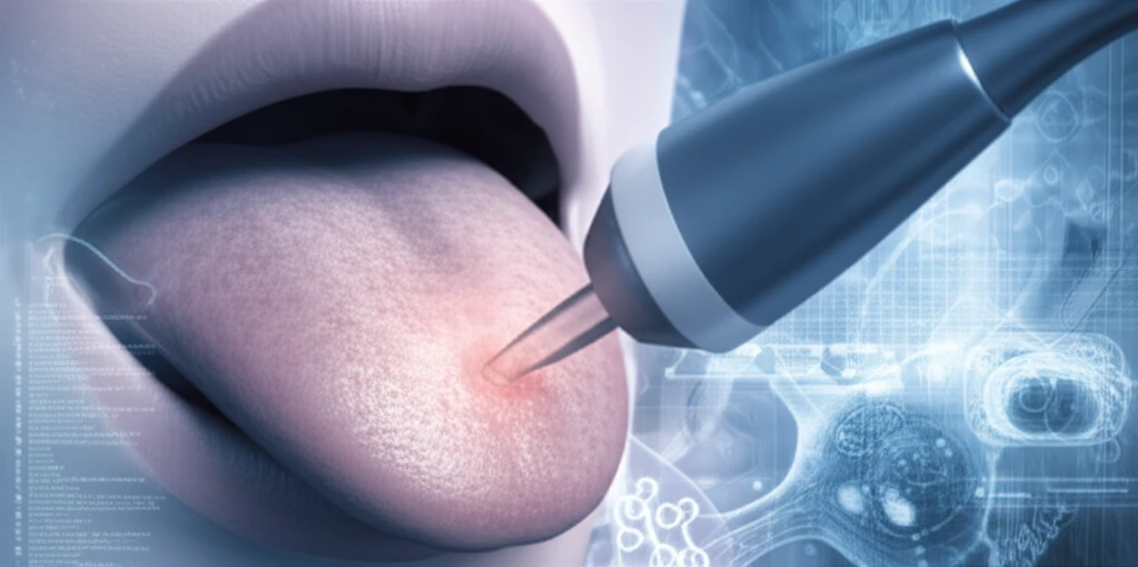
How Does Strain Elastography Work?

Illustration of ultrasound probe examining a tongue lesion.

Strain elastography is based on the principle that cancerous tissues are generally stiffer than normal tissues. The technique involves applying slight pressure to the tissue using an ultrasound probe and measuring the tissue's deformation. This information is then used to create a visual representation of tissue stiffness.

In this study, researchers used a linear 14 MHz transducer to perform strain elastography on two patients with squamous cell carcinoma (SCC) of the tongue. The procedure involved:

  • Applying gentle compression and decompression to the tongue using the ultrasound probe.
  • Monitoring tissue strain over time using a strain graph.
  • Using integrated software to measure quantitative strain (%) within regions of interest (ROIs).
  • Comparing strain measurements between normal tissues and lesions.
The study found that the average strain in normal tongue tissue was significantly higher than in tongue SCC, indicating that cancerous tissues were indeed stiffer. Specifically, in one patient, the average strain of normal tissue was 1.468% compared to 0.000% in the SCC. Similar results were observed in the second patient, with normal tissue showing a strain of 1.007% and SCC showing 0.000%.

The Future of Tongue Cancer Diagnosis

This preliminary study suggests that strain elastography using intraoral ultrasonography is a promising technique for characterizing and differentiating normal tissues and SCC in the tongue. While the study involved only two patients, the results warrant further investigation with larger sample sizes.

The potential benefits of this approach include:

<ul><li>Non-invasive: It does not require surgical biopsies.</li><li>Relatively inexpensive: It utilizes existing ultrasound technology with specialized software.</li><li>Easy to use: The procedure can be performed by trained clinicians.</li><li>Early detection: It may help identify cancerous lesions at an earlier stage.</li></ul>

About this Article -

This article was crafted using a human-AI hybrid and collaborative approach. AI assisted our team with initial drafting, research insights, identifying key questions, and image generation. Our human editors guided topic selection, defined the angle, structured the content, ensured factual accuracy and relevance, refined the tone, and conducted thorough editing to deliver helpful, high-quality information.See our About page for more information.

This article is based on research published under:

DOI-LINK: 10.5624/isd.2018.48.1.45, Alternate LINK

Title: Strain Elastography Of Tongue Carcinoma Using Intraoral Ultrasonography: A Preliminary Study To Characterize Normal Tissues And Lesions

Subject: Radiology, Nuclear Medicine and imaging

Journal: Imaging Science in Dentistry

Publisher: Korean Academy of Oral and Maxillofacial Radiology

Authors: Ichiro Ogura, Yoshihiko Sasaki, Mikiko Sue, Takaaki Oda

Published: 2018-01-01

Everything You Need To Know

1

What is Intraoral ultrasonography and Strain elastography?

Intraoral ultrasonography is a type of imaging method used to visualize the inside of the mouth, including the tongue. It can help to identify lesions. Strain elastography is a technique used with Intraoral ultrasonography that assesses the stiffness of tissues, potentially offering additional insights into the nature of tongue lesions. Together, they provide a less invasive way to characterize tongue lesions, which could improve the detection and characterization of tongue carcinoma. The combination aims to offer a promising avenue for early diagnosis, which is crucial for successful treatment. It allows for the differentiation between normal tissues and cancerous lesions, which is challenging with traditional methods like visual exams and palpation.

2

Why is Strain elastography important in the context of tongue cancer diagnosis?

Strain elastography is significant because it can potentially improve the accuracy of diagnosing tongue cancer. Cancerous tissues tend to be stiffer than normal tissues, and Strain elastography measures this difference in stiffness. By using Intraoral ultrasonography with strain elastography, doctors can gain additional insights into the nature of tongue lesions. This is important because early detection of tongue cancer is crucial for effective treatment. The technique assesses tissue deformation to create a visual representation of tissue stiffness, which can help distinguish between benign and cancerous lesions.

3

How was Strain elastography used in the study?

The study utilized Intraoral ultrasonography combined with Strain elastography by using a linear 14 MHz transducer to perform strain elastography on two patients with squamous cell carcinoma (SCC) of the tongue. The procedure involved applying gentle compression and decompression to the tongue using the ultrasound probe, monitoring tissue strain over time using a strain graph, using integrated software to measure quantitative strain (%) within regions of interest (ROIs), and comparing strain measurements between normal tissues and lesions. The researchers were able to measure the strain in the normal tongue tissue and cancerous lesions and compare them.

4

What is the significance of the findings related to Squamous cell carcinoma (SCC) and normal tissue strain?

Squamous cell carcinoma (SCC) is a type of cancer that can affect the tongue. The study found that the average strain in normal tongue tissue was significantly higher than in tongue SCC, indicating that cancerous tissues were indeed stiffer. In one patient, the average strain of normal tissue was 1.468% compared to 0.000% in the SCC. Similar results were observed in the second patient, with normal tissue showing a strain of 1.007% and SCC showing 0.000%. This means that the cancerous tissues showed less deformation under pressure compared to normal tissue, confirming the principle that cancerous tissues are generally stiffer. This finding is important because it supports the potential of Strain elastography, in conjunction with Intraoral ultrasonography, to distinguish between cancerous and normal tissues.

5

What are the implications of this study for the future of tongue cancer diagnosis?

While the study only involved two patients, the results suggest that Strain elastography, when used with Intraoral ultrasonography, is a promising technique for the early detection and characterization of tongue cancer. The implication is that this combined approach could lead to earlier and more accurate diagnoses. This preliminary research aims to determine if this combined approach can differentiate between normal tissues and cancerous lesions in the tongue, potentially leading to earlier and more accurate diagnoses. Further studies with larger sample sizes are needed to confirm these findings and assess the full potential of this technique. If successful, it could revolutionize how tongue cancer is diagnosed, offering a less invasive and more effective diagnostic tool.

Newsletter Subscribe

Subscribe to get the latest articles and insights directly in your inbox.