Illustration of nanoimaging with soft X-ray and EUV lasers inside a cell, highlighting nanoscale details.

Tiny Tech, Big Discoveries: How Nanoimaging is Revolutionizing Biology and Beyond

"Unlocking the Secrets Within: Exploring the Revolutionary World of Nanoimaging Using Soft X-ray and EUV Laser-Plasma Sources."


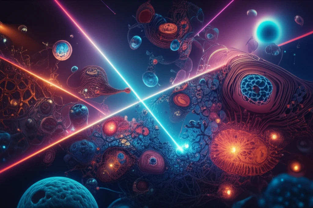
In the ever-evolving landscape of scientific discovery, the ability to see smaller and understand better is paramount. We stand at the cusp of a new era, thanks to advancements in nanoimaging technologies. These technologies, which use extreme ultraviolet (EUV) and soft X-ray (SXR) microscopy, are helping us to observe and analyze objects at the nanometer scale.

Imagine peering into the inner workings of a cell or examining the intricate structures of a new material, all with unprecedented detail. This is now possible, thanks to the development of compact and powerful EUV and SXR sources, especially those based on laser-plasma technology. These tools are not just enhancing our vision; they're reshaping the boundaries of what we can explore and understand.

This article delves into the fascinating world of nanoimaging, focusing on the latest advancements in EUV and SXR laser-plasma sources. We'll explore how these technologies work, their applications in fields ranging from biology to materials science, and how they are paving the way for future discoveries.

Peering into the Nano World: The Science Behind Nanoimaging

Illustration of nanoimaging with soft X-ray and EUV lasers inside a cell, highlighting nanoscale details.

At the heart of nanoimaging lies the use of extremely short-wavelength radiation, specifically EUV and SXR. These wavelengths are absorbed differently by various materials, creating a high contrast that allows us to distinguish between different components of a sample. For example, SXR is particularly useful for biological imaging because of the contrast it provides between carbon and water, the main elements of biological matter. This contrast helps scientists to visualize and understand biological structures with remarkable clarity.

The traditional methods for nanoimaging often involve large-scale facilities like synchrotrons and free-electron lasers, which are expensive and have limited accessibility. However, recent developments have led to the creation of compact, desk-top EUV and SXR sources, particularly those using laser-plasma technology. These sources use a high-powered laser to create a plasma, which then emits the desired EUV or SXR radiation. This approach allows researchers to conduct experiments in their own laboratories, accelerating the pace of discovery.

  • Laser-Plasma Sources: These sources use high-powered lasers to generate plasma, which emits EUV and SXR radiation.
  • Water Window: This spectral range (2.3–4.4 nm) is ideal for biological imaging due to the contrast between carbon and water.
  • Compact Microscopes: Desk-top systems offer an accessible alternative to large-scale facilities.
  • Applications: Nanoimaging is used in biology, material science, and nanotechnology.
The development of these compact systems is crucial, and efforts are being made to improve spatial resolution and decrease exposure times. Moreover, recent breakthroughs have enabled researchers to visualize the internal structure of cells and nanostructures with resolutions down to a few tens of nanometers. These advancements are not just about making things smaller; they're about seeing more clearly and understanding the intricate details that drive the world around us.

The Future of Seeing: Nanoimaging and Beyond

As nanoimaging technologies continue to evolve, the future is brimming with potential. From revolutionizing medical diagnostics to enabling the creation of novel materials, the impact of EUV and SXR microscopy will only continue to grow. The accessibility and advancements in this area promise to democratize scientific research, enabling a wider range of researchers to contribute to this exciting field. By opening new windows into the nano world, we're not just seeing smaller; we're understanding more, creating more, and ultimately, changing the world.

About this Article -

This article was crafted using a human-AI hybrid and collaborative approach. AI assisted our team with initial drafting, research insights, identifying key questions, and image generation. Our human editors guided topic selection, defined the angle, structured the content, ensured factual accuracy and relevance, refined the tone, and conducted thorough editing to deliver helpful, high-quality information.See our About page for more information.

This article is based on research published under:

DOI-LINK: 10.1051/epjconf/201816703001, Alternate LINK

Title: Nanoimaging Using Soft X-Ray And Euv Laser-Plasma Sources

Subject: General Medicine

Journal: EPJ Web of Conferences

Publisher: EDP Sciences

Authors: Przemyslaw Wachulak, Alfio Torrisi, Mesfin Ayele, Andrzej Bartnik, Joanna Czwartos, Łukasz Węgrzyński, Tomasz Fok, Henryk Fiedorowicz

Published: 2018-01-01

Everything You Need To Know

1

What makes nanoimaging using soft X-rays and EUV laser-plasma sources so revolutionary for biological studies?

Nanoimaging with soft X-rays (SXR) and EUV laser-plasma sources is revolutionary for biology because of the high contrast it provides between carbon and water, the primary elements in biological matter. This 'water window' allows scientists to visualize cellular structures with remarkable clarity, enabling detailed studies of cells and tissues at the nanometer scale without the need for staining or invasive preparation methods. This level of detail was previously unattainable with traditional microscopy techniques, which often require specialized sample preparation that can alter or damage the specimens.

2

How do compact, desk-top EUV and SXR sources, particularly those using laser-plasma technology, differ from traditional nanoimaging methods, and what are the implications of these differences?

Traditional nanoimaging methods often rely on large-scale facilities like synchrotrons and free-electron lasers, which are expensive and have limited accessibility. Compact, desk-top EUV and SXR sources, especially those based on laser-plasma technology, offer a more accessible alternative. These sources use high-powered lasers to generate plasma, which emits EUV or SXR radiation. This allows researchers to conduct experiments in their own laboratories, accelerating the pace of discovery and democratizing scientific research. This localized approach reduces the need for travel and extensive collaboration, fostering innovation and making advanced imaging techniques available to a wider range of researchers and institutions.

3

What are the primary applications of nanoimaging beyond biology, and how do EUV and SXR laser-plasma sources contribute to these advancements?

Beyond biology, nanoimaging finds applications in materials science and nanotechnology. In materials science, it allows researchers to examine the intricate structures of new materials at the nanometer scale, aiding in the development of advanced materials with tailored properties. In nanotechnology, it enables the visualization and analysis of nanostructures, facilitating the creation of novel devices and systems. EUV and SXR laser-plasma sources are crucial in these advancements by providing the necessary high-resolution imaging capabilities in a more accessible and cost-effective manner, accelerating research and development in these fields.

4

What is the 'water window' in the context of nanoimaging, and why is it important for biological applications?

The 'water window' refers to the spectral range of 2.3–4.4 nm in the electromagnetic spectrum. This range is particularly important for biological applications of nanoimaging because water and carbon absorb radiation differently, resulting in high contrast. Because water is the main constituent, the water window allows scientists to visualize biological structures with clarity. This is essential for detailed studies of cells and tissues at the nanometer scale, enabling researchers to observe and analyze biological processes without the need for staining or invasive preparation methods that could alter the natural state of the sample.

5

How is the development of nanoimaging technologies, specifically EUV and SXR microscopy, expected to impact future medical diagnostics and the creation of new materials?

Advancements in nanoimaging technologies, particularly EUV and SXR microscopy, are expected to revolutionize medical diagnostics by enabling earlier and more accurate detection of diseases at the cellular and molecular levels. This could lead to personalized medicine approaches, where treatments are tailored to an individual's specific condition based on detailed nanoimaging analysis. In materials science, these technologies will facilitate the creation of novel materials with enhanced properties by allowing researchers to visualize and manipulate structures at the nanoscale. This could lead to breakthroughs in areas such as energy storage, advanced electronics, and high-performance materials with applications across various industries.

Newsletter Subscribe

Subscribe to get the latest articles and insights directly in your inbox.