Thyroid Scans vs. Biopsy: Which Test Is Best for Spotting Cancer?
"A new study explores how combining thyroid scans can improve accuracy in detecting malignant thyroid nodules, potentially reducing the need for unnecessary biopsies."
The journey of diagnosing thyroid nodules can be filled with uncertainty. While most thyroid nodules are benign, the possibility of malignancy always looms, leading to anxiety and the need for accurate diagnostic tools. Traditionally, fine needle aspiration cytology (FNAC) has been a cornerstone in evaluating these nodules, but its limitations in certain cases have prompted the search for more reliable, non-invasive methods.
Thyroid scintigraphy, or thyroid scanning, has been used to assess thyroid nodules, but questions remain regarding its role. Although critical for determination of autonomously functioning thyroid tissue, conventional scanning has limitations in distinguishing between benign and malignant nodules. Researchers have explored various radiopharmaceuticals to improve the accuracy of thyroid imaging.
This article delves into a study comparing conventional thyroid scintigraphy with newer techniques using 99mTc tetrofosmin (Myoview) and 99mTc (V) DMSA, combined with FNAC, to evaluate solitary thyroid nodules. We'll explore how these methods can improve the accuracy of diagnosis and potentially reduce the need for invasive procedures.
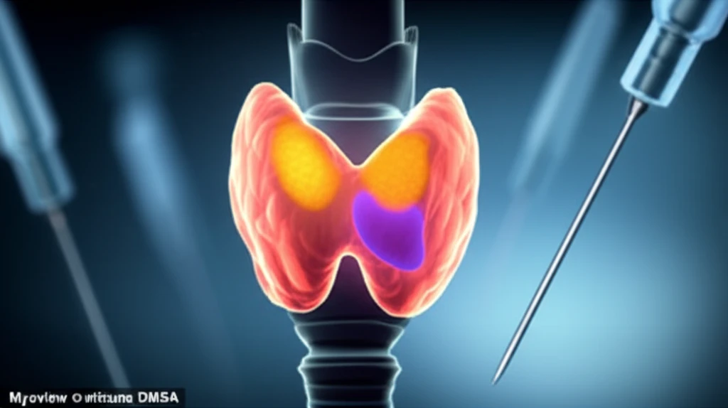
Myoview and DMSA: A Combined Approach to Thyroid Nodule Evaluation
The study involved 103 patients, predominantly women, with solitary thyroid nodules. All patients underwent a comprehensive evaluation, including a detailed medical history, physical examination, thyroid function tests, and various thyroid scans, including conventional 99mTc pertechnetate scans, 99mTc tetrofosmin (Myoview) scans, and 99mTc (V) DMSA scans. Cytologic assessments were also performed on thyroid nodules that appeared "cold" on conventional scans.
- Conventional 99mTc pertechnetate scans: These scans had limited value in distinguishing between benign and malignant nodules.
- Early-phase Myoview scans: High uptake in the early phase was observed in both malignant and benign nodules, indicating it's not specific for malignancy.
- Combined Myoview and DMSA scans: Researchers found that combining Myoview and DMSA scans provided valuable diagnostic information. Absence of tracer accumulation in both scans strongly suggested a benign nodule.
- Quantitative Analysis: High retention index of Myoview in malignant lesions compared to benign lesions suggested malignancy. A cut-off point >14% could separate between benign and malignant lesions.
Improving Thyroid Nodule Assessment: The Future of Diagnosis
The study suggests that Myoview uptake in cold nodules is more indicative of the aggressiveness or grade of a malignant tumor rather than simply distinguishing between benign and malignant lesions. To differentiate between these two, both Myoview and DMSA scans, combined with a regression equation, are needed.
This research highlights the potential of combining different imaging techniques to improve diagnostic accuracy. The use of Myoview and DMSA scans can offer a less invasive route to identifying potential thyroid cancers, and refining the selection process for FNAC, ensuring that biopsies are performed when they are most likely to provide valuable information.
Future studies should explore the findings and refine the diagnostic algorithms to improve the detection of thyroid malignancies while minimizing unnecessary invasive procedures, ultimately leading to better patient outcomes and reduced anxiety for individuals with thyroid nodules.