
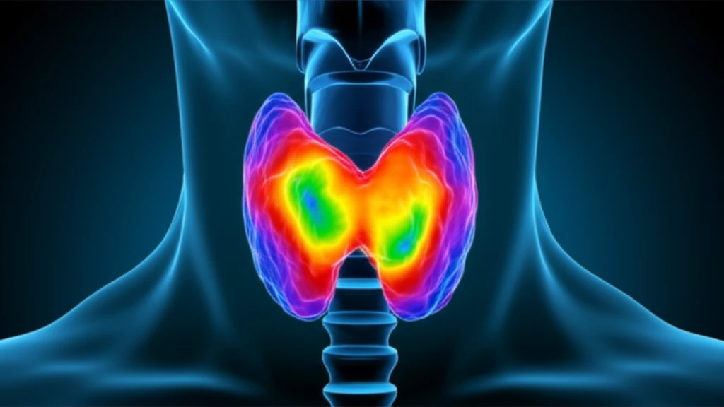
Thyroid Cancer Detection: Can Advanced MRI Scans Improve Accuracy?

"Discover how Diffusion-Weighted MRI (DW-MRI) enhances thyroid nodule assessment, potentially reducing unnecessary surgeries."
Thyroid nodules are common, affecting a significant portion of the population. While most are benign, the possibility of malignancy always looms, creating anxiety and diagnostic challenges. Current methods, like ultrasound and fine-needle aspiration (FNA) biopsies, have limitations in definitively ruling out cancer, leading to potential overtreatment and unnecessary surgeries.
Advanced imaging techniques offer promising solutions. Diffusion-Weighted Magnetic Resonance Imaging (DW-MRI) is emerging as a valuable tool for assessing thyroid nodules. DW-MRI measures the movement of water molecules within tissues, providing insights into cellular density and integrity – characteristics that differ significantly between benign and malignant tissues.
This article explores how DW-MRI, particularly when combined with Apparent Diffusion Coefficient (ADC) values, can improve the accuracy of thyroid cancer detection. We'll delve into the research, the benefits, and what this means for individuals facing the uncertainty of thyroid nodules.
Current Diagnostic Landscape
Routine thyroid-nodule evaluation commonly includes ultrasonography, thyroid scintigraphy, positron emission tomography-computed tomography, and fine-needle biopsy. The listed research reports that conventional MRI has limited value for differentiating thyroid nodules, while MRI has become a beneficial modality in thyroid diseases. Multi-parameter diffusion-weighted imaging is being investigated for quantitative evaluation of thyroid nodules.
Understanding Diffusion-Weighted MRI (DW-MRI) and ADC Values
DW-MRI works by detecting how water molecules move within body tissues. In areas where cells are densely packed (like in tumors), water movement is restricted. This restriction is measured by the Apparent Diffusion Coefficient (ADC). Malignant tissues typically show lower ADC values compared to benign tissues because of their higher cellular density.
- How DW-MRI Works: Measures water molecule movement in tissues.
- ADC Values: Quantify water diffusion; lower values suggest malignancy.
- Cellular Density: Malignant tissues have higher cellular density, restricting water movement.
Recent MRI Evidence
A systematic review describes thyroid nodules as highly prevalent, affecting up to 75% of the population, although most are benign. Diffusion-weighted MRI evaluates tissue characteristics through the apparent diffusion coefficient, and researchers are studying it as an imaging-based classification method. In a separate comparison, adenomatous nodules had a higher APTw value than papillary thyroid carcinomas, measuring 3.3 ± 1.3% versus 1.8 ± 0.7%, respectively.
Limits and Uncertainty
The available evidence does not establish diffusion-weighted MRI as a replacement for definitive diagnosis. One source states that surgery remains the gold standard for distinguishing benign from malignant thyroid nodules, although it can be invasive and unnecessary in indeterminate cases. Another meta-analysis describes DWI as promising but emphasizes that its accuracy remains an important issue; the 2025 analysis included 46 studies.
DWI Versus Current Workups
The source reports that limited specificity in current diagnostic approaches can contribute to unnecessary interventions and overtreatment. Diffusion-weighted MRI is presented as a non-invasive method that quantifies tissue microstructure using the apparent diffusion coefficient. Its proposed role is thyroid-nodule classification, rather than an established replacement for existing diagnostic pathways.
The Future of Thyroid Cancer Diagnostics
DW-MRI holds considerable promise as a supplementary tool in the diagnostic process for thyroid nodules. By providing a non-invasive method to assess the characteristics of thyroid nodules, DW-MRI can assist clinicians in making more informed decisions about the need for biopsy or surgery. While further research is needed to refine the technique and establish standardized protocols, DW-MRI is paving the way for more accurate and personalized approaches to thyroid cancer detection.
Potential Clinical Role
Researchers have tested diffusion-weighted MRI as an advanced approach for evaluating thyroid nodules. The cited study reports differences between benign and suspicious nodules, while noting that ultrasound and cell sampling can leave some nodules unclear. These findings suggest that DW-MRI may add information to existing evaluation, but the source presents it as an investigated tool rather than a confirmed diagnostic replacement.
Technical and Treatment Considerations
Surgery remains the gold standard for distinguishing benign from malignant thyroid nodules, but the cited source notes that it is invasive and often unnecessary for indeterminate cases. Diffusion-weighted MRI may help distinguish benign from suspicious nodules and support treatment decisions, potentially reducing unnecessary surgery. However, the accuracy of DW-MRI is affected by technical acquisition parameters, whose impact remains unclear.