The Future is Now: How Nanofiber Scaffolds Are Revolutionizing Cardiovascular Implants
"Explore the groundbreaking research into nanofibrous polyamide 6 scaffolds and their potential to transform the success rates of cardiovascular implants, paving the way for more biocompatible solutions."
The field of implant materials is constantly evolving, with researchers seeking innovative ways to improve the integration and functionality of synthetic devices within the human body. One promising avenue is the use of synthetic scaffolds, which provide a framework for tissue regeneration and cell growth. These scaffolds aim to mimic the natural extracellular matrix, supporting cells as they rebuild damaged or diseased tissues.
Cardiovascular implants, in particular, face significant challenges related to biocompatibility. The risk of thrombus formation (blood clots) and neointimal hyperplasia (thickening of the artery wall) can lead to implant failure and further complications for patients. Traditional implants often struggle to promote effective endothelialization, the process where a layer of endothelial cells forms on the implant surface, creating a natural, anti-thrombotic barrier.
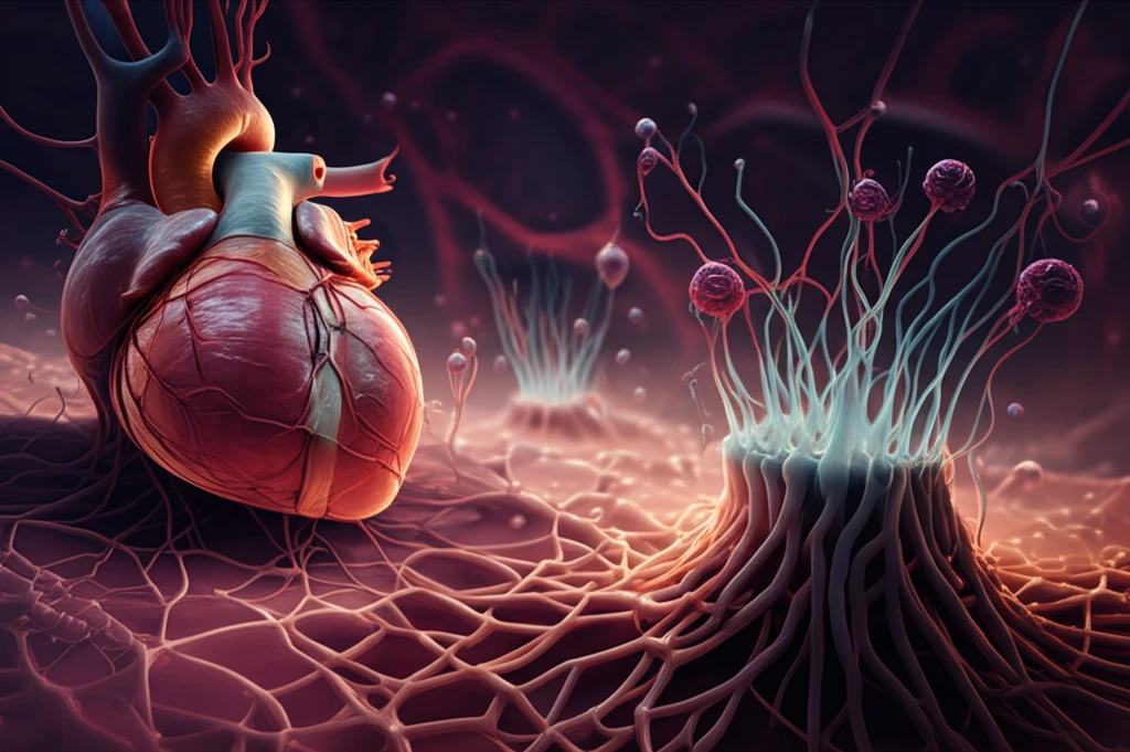
Recent studies have focused on nanofiber technology as a potential solution to these challenges. Nanofibers, with their ultra-fine structure, closely resemble the natural extracellular matrix, offering an ideal environment for cell attachment and growth. A groundbreaking study investigated the use of nanofibrous polyamide 6 (PA-6) scaffolds, exploring their ability to enhance endothelial cell adhesion and overall biocompatibility. This article delves into the fascinating world of nanofiber scaffolds and their potential to revolutionize cardiovascular implants.
Nanofiber Scaffolds: A New Hope for Cardiovascular Implants
The core of this innovative approach lies in electrospinning, a technique used to create nanofibrous nonwovens of polyamide 6 (PA-6). Researchers meticulously characterized these scaffolds, assessing their mechanical strength and biological performance. The mechanical strength was evaluated through uniaxial tensile testing, while biological performance was gauged by measuring cell viability and analyzing cellular morphology using human umbilical vein endothelial cells (EA.hy926) and human fibroblasts (HT-1080).
- Selective Cell Adhesion: Nanofiber scaffolds promoted endothelial cell adhesion over fibroblast adhesion.
- Excellent Biocompatibility: All tested materials showed high cell viability.
- Mechanical Strength: The scaffolds demonstrated robust mechanical properties suitable for cardiovascular applications.
- Material Innovation: Utilized polyamide 6 (PA-6), a biocompatible polymer.
The Path Forward: Refining Nanofiber Technology for Clinical Use
While the study's findings are highly encouraging, further research is needed to translate these advancements into clinical applications. Future studies should focus on optimizing the nanofiber scaffold design to achieve even greater control over cell adhesion and tissue regeneration. Additionally, long-term studies are necessary to evaluate the durability and performance of these implants in vivo. By continuing to explore the potential of nanofiber technology, we can pave the way for cardiovascular implants that offer improved biocompatibility, reduced complications, and ultimately, better outcomes for patients.