
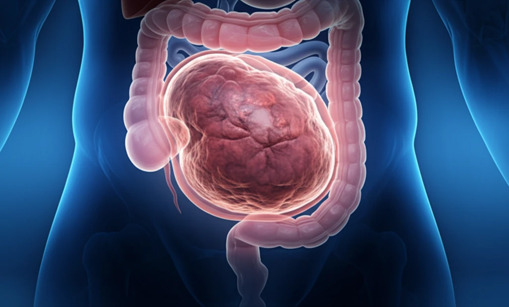
The Enigmatic Mesothelial Cyst: Unraveling a Rare Abdominal Mimic

"A deep dive into a rare case where a simple mesothelial cyst masquerades as a hydatid cyst, challenging diagnostic assumptions."
In the realm of medical anomalies, few cases are as perplexing as when a benign condition mimics a more concerning ailment. Mesothelial cysts, simple and often overlooked, present a diagnostic puzzle when they imitate other, more well-known conditions. This article delves into a fascinating case where a mesothelial cyst cleverly imitated a hydatid cyst, leading to a diagnostic journey that underscores the importance of thorough investigation and a nuanced understanding of rare conditions.
Hydatid cysts, typically caused by the Echinococcus parasite, are commonly found in the liver and lungs. Their presence often necessitates careful management due to potential complications. However, when a non-parasitic cyst presents similarly, the diagnostic pathway becomes less clear-cut. This is precisely what occurred in the case highlighted in the Pan African Medical Journal, where a 55-year-old female was initially suspected of harboring a hydatid cyst in her liver, only for further investigation to reveal a mesothelial cyst.
The case not only illuminates the challenges in distinguishing between different types of abdominal cysts but also emphasizes the critical role of advanced imaging and pathological examination in arriving at an accurate diagnosis. By exploring this specific instance, we gain insights into the nature of mesothelial cysts, their clinical presentation, and the strategies used to differentiate them from other abdominal abnormalities.
An Extremely Rare Condition
Mesothelial cysts are extremely rare benign fluid-filled sacs lined by mesothelial cells that arise from the mesothelium lining the body's cavities, including the chest, abdomen, and pelvis. They are also referred to as primary retroperitoneal cysts of mesothelial origin, and only a limited number of confirmed cases have been reported in the medical literature. Diaphragmatic mesothelial cysts are classified as rare congenital lesions, and historically, only a small number of patients—mostly adults—have been documented undergoing surgical treatment for them.
Diagnostic Ambiguity and Classification Challenges
Peritoneal mesothelial cysts present a clinical dilemma due to uncertainty surrounding their true pathogenic nature. Multiple overlapping definitions and terminology have been applied to these lesions, including 'benign multicystic mesothelioma,' 'cystic mesothelioma,' and 'multilocular peritoneal inclusion cyst,' which complicates consistent diagnosis and management. Histopathological classification schemes have been proposed to organize cysts by their features and origin, but the heterogeneity of these entities means that no single, universally accepted diagnostic framework has been firmly established.
Early Documentation and Pathological Foundations
A peritoneal simple mesothelial cyst was historically recognized as a very rare mesenteric cyst of mesothelial origin, with lesion size typically ranging from a few centimeters up to approximately 10 cm. Cysts of mesothelial origin were identified as rare lesions observed more frequently in young and middle-aged women, predominantly benign and located in the mesenteries or omentum, with diagnosis based on clinical examination and radiographic imaging. Early case reports, including those of benign cystic mesothelioma in the mediastinum, established that mesothelial cysts encompass a group of lesions defined as pleuropericardial cysts, lymphogenous cysts, pleural cysts, or simple mesothelial cysts, often discovered incidentally on radiological examinations.
The Case Unveiled: A Diagnostic Odyssey
The patient, a 55-year-old woman, presented with right hypochondrium pain, leading to an initial suspicion of a liver hydatid cyst. An ultrasound revealed a 14 cm cyst, classified as Type 2 according to Gharbi's classification, in the right lobe of her liver. This initial finding strongly suggested a hydatid cyst, a parasitic condition endemic in many regions. However, further investigation via abdominal scanning uncovered more than initially suspected.
- The liver cyst was indeed treated conservatively, aligning with hydatid cyst management protocols.
- A right colo-parietal detachment revealed a retroperitoneal cyst with thin, translucent walls and serous content.
- Enucleation of this cyst was performed, allowing for detailed pathological examination.
- Post-operative pathological analysis confirmed the cyst to be a simple mesothelial cyst, altering the course of treatment and follow-up.
Emerging Case Reports and Site-Specific Findings
Recent literature continues to document mesothelial cysts arising in diverse anatomical locations, including the spleen, where they are described as developmental disorders characterized by unilocular morphology with smooth, well-defined margins. Mesothelial cysts of the liver round ligament are recognized as rare entities, with approximately five cases identified in the literature, and reported diameters ranging from 5 to 14 cm. Uterine mesothelial cysts represent a particular diagnostic challenge due to their low incidence, with very few cases reported in the English-language medical literature, as highlighted by recent case reports in young nulliparous patients.
Controversies in Treatment and Limited Data
Intrathoracic mesothelial cysts are understood as congenital lesions resulting from abnormal development of the pericardial coelom, yet the preferred treatment for giant mediastinal mesothelial cysts remains a controversial topic with no definitive consensus. Mesothelial cysts are considered rare overall, with limited data available on their true prevalence and incidence across populations. While they can occur in individuals of any age, they are more commonly diagnosed in adults, and although there is no clear gender predilection, some studies suggest a slight female predominance that has not been conclusively established.
Distinguishing Mesothelial Cysts from Mimics
Mesenteric cysts and cystic lymphangiomas are scarce entities that are clinically and radiologically almost identical, yet their histological structures are fundamentally different, making accurate differentiation essential. Mesothelial cysts arise from the mesothelial cells lining the peritoneum and are usually small and asymptomatic, whereas enteric cysts originate from the intestines, representing a distinct pathological category. The radiological overlap between mesothelial cysts and other cystic abdominal lesions underscores the importance of histopathological confirmation for accurate diagnosis.
Lessons Learned and Clinical Implications
This case underscores several critical points for clinicians. First, it highlights the importance of maintaining a broad differential diagnosis when evaluating abdominal cysts. While hydatid cysts are relatively common in certain regions, other cystic lesions, such as mesothelial cysts, should also be considered, especially when imaging characteristics are atypical. Second, advanced imaging techniques, such as CT scans with contrast, can provide valuable information about the nature of cysts, including their location, content, and enhancement patterns. Finally, pathological examination remains the gold standard for definitive diagnosis, particularly when differentiating between benign and potentially more serious conditions. By remaining vigilant and employing a comprehensive diagnostic approach, clinicians can ensure accurate diagnoses and optimal patient outcomes.
Diagnostic Precision Remains Elusive
Mesothelial inclusion cyst, sometimes termed benign cystic mesothelioma, is a rare benign tumor with only approximately 130 cases reported in the literature as of the most recent comprehensive clinicopathological analysis. Experts have noted that accurate diagnosis and optimal management of this condition remain uncertain, reflecting the challenges posed by the entity's rarity and variable presentation. The persistent diagnostic ambiguity highlights the need for greater awareness among clinicians and pathologists to avoid mischaracterization of these lesions.
Expanding the Literature on Rare Presentations
Intramyometrial mesothelial cysts of the uterus are described as extremely rare, with only a handful of cases documented in the medical literature, and recent case reports aim to further characterize their clinical, radiological, and histopathological features. Additionally, mesothelial cysts must be considered in the differential diagnosis of scrotal and testicular masses, as they can be confused with testicular tumors, potentially leading to unnecessary surgical interventions. Continued reporting of rare cases across diverse anatomical sites will be essential for building a more comprehensive understanding of mesothelial cyst biology and optimal clinical management.
Navigating Rarity in Clinical Practice
Mesothelial cysts exist at the intersection of several systemic challenges in medicine: their extreme rarity means that most clinicians may encounter only a handful of cases, if any, throughout their careers, making pattern recognition and accumulated experience difficult to achieve. The lack of large-scale epidemiological data hampers the development of evidence-based screening protocols or standardized management pathways. Addressing these gaps will likely require multicenter collaboration and the creation of centralized registries to aggregate data on this under-studied condition.
Patient Experience and Clinical Uncertainty
For patients diagnosed with a mesothelial cyst, the journey from symptom onset to definitive diagnosis can be fraught with anxiety, particularly when imaging findings are ambiguous and the possibility of malignancy must be excluded. The rarity of the condition means that patients may be referred to multiple specialists before arriving at a correct diagnosis, adding emotional and financial burden. Improving clinician awareness and developing clearer diagnostic algorithms could meaningfully reduce this burden and accelerate time to appropriate care.