Illustration of duplicated axillary arch muscles.

The Curious Case of the Duplicated Axillary Arch: What It Means for Your Body

"Unveiling a Rare Muscular Variation and Its Potential Impact on Shoulder Health and Surgical Outcomes"


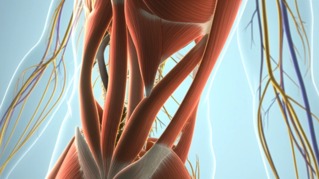
The axillary region, nestled between the neck and upper limb, is a bustling hub of nerves, blood vessels, and lymph nodes. Its complex anatomy means that variations in muscle structure can sometimes occur, potentially leading to complications.

One such variation is the axillary arch muscle, also known as Langer's muscle. This muscular slip, arising from the latissimus dorsi, can sometimes obstruct vessels and nerves, contributing to conditions like thoracic outlet syndrome or shoulder instability. It can even impact surgical interventions, such as breast surgery, and lymphatic drainage.

Recent research has uncovered an even rarer occurrence: a duplicated axillary arch muscle. This article delves into this unique anatomical finding, exploring its potential implications for your health and well-being.

Duplicated Axillary Arch: A Closer Look at This Rare Variation

Illustration of duplicated axillary arch muscles.

A recent anatomical study detailed the discovery of a duplicated axillary arch muscle during a routine dissection. In a 33-year-old male cadaver, researchers identified two distinct muscular slips originating from the same inferolateral border of the latissimus dorsi muscle on the right side.

These duplicated muscles differed in length and insertion points:

  • Long Slip: Approximately 13.5 cm long, inserting into the aponeurosis of the pectoralis major (the large chest muscle).
  • Short Slip: About 9.1 cm long, inserting into the deep fascia covering the pectoralis minor (a smaller chest muscle located beneath the pectoralis major).
While both slips originated from the latissimus dorsi near the second and third ribs, they had distinct insertion points. Interestingly, researchers didn't find separate nerve supplies for each slip, but noted small branches of intercostobrachial nerves nearby. The thoracodorsal vessels and nerve, lateral thoracic vessels, and long thoracic nerve were located underneath the muscular slips.

What Does This Mean for You? Implications and Considerations

While this duplicated axillary arch is a rare finding, understanding its potential implications is crucial. The presence of such muscular variations can contribute to:

<ul><li><b>Nerve Compression:</b> Axillary arch muscles may compress nearby nerves like the axillary vein, musculocutaneous, median, and ulnar nerves, potentially causing pain, numbness, or weakness in the arm and hand.</li><li><b>Limited Shoulder Movement:</b> The presence of an axillary arch can restrict shoulder elevation and hyperabduction, impacting range of motion.</li><li><b>Surgical Complications:</b> Surgeons need to be aware of this variation before performing procedures in the axillary region, such as axillary lymphadenectomy (lymph node removal) for breast cancer treatment. The muscle can obstruct the surgical field or be damaged during the procedure.</li></ul>

Although this specific study couldn't assess functional disorders due to the use of a cadaver, it highlights the importance of anatomical awareness. If you experience unexplained shoulder pain, limited movement, or neurological symptoms in your arm, it's essential to consult with a healthcare professional. Knowledge of anatomical variations like the duplicated axillary arch can aid in accurate diagnosis and treatment.

About this Article -

This article was crafted using a human-AI hybrid and collaborative approach. AI assisted our team with initial drafting, research insights, identifying key questions, and image generation. Our human editors guided topic selection, defined the angle, structured the content, ensured factual accuracy and relevance, refined the tone, and conducted thorough editing to deliver helpful, high-quality information.See our About page for more information.

This article is based on research published under:

DOI-LINK: 10.5115/acb.2012.45.4.288, Alternate LINK

Title: Duplicated Axillary Arch Muscles Arising From The Latissimus Dorsi

Subject: Cell Biology

Journal: Anatomy & Cell Biology

Publisher: Korean Association of Anatomists

Authors: Sitthichai Iamsaard, Nongnut Uabundit, Kimaporn Khamanarong, Kittisak Sripanidkulchai, Kowit Chaiciwamongkol, Malivalaya Namking, Somsiri Ratanasuwan, Porntip Boonruangsri, Wiphawi Hipkaeo

Published: 2012-01-01

Everything You Need To Know

1

What exactly is a duplicated axillary arch muscle?

The axillary arch muscle, specifically a duplicated one, is a rare anatomical variation where two distinct muscular slips originate from the latissimus dorsi. In the case described, one slip (the long slip) inserted into the aponeurosis of the pectoralis major, while the other (the short slip) inserted into the deep fascia covering the pectoralis minor. This variation could potentially affect shoulder movement and nerve function.

2

How could a duplicated axillary arch muscle impact surgical procedures?

The duplicated axillary arch muscle, being a variation in the muscular structure of the axillary region, has the potential to impact surgical procedures, particularly those involving the breast or axilla. Surgeons need to be aware of this variation to avoid accidental damage to nerves and vessels that may be compressed or displaced by the duplicated muscle slips. The thoracodorsal vessels and nerve, lateral thoracic vessels, and long thoracic nerve were located underneath the muscular slips.

3

What are the potential symptoms or health issues associated with having a duplicated axillary arch muscle?

While no direct symptoms are explicitly linked to a duplicated axillary arch muscle, the presence of any axillary arch variation, including a duplicated one, could potentially contribute to conditions like thoracic outlet syndrome or shoulder instability if it obstructs vessels or nerves in the axillary region. The duplicated muscle might affect lymphatic drainage. However, most people with this variation might be asymptomatic.

4

Where was the duplicated axillary arch muscle found and described?

The study referenced found a duplicated axillary arch muscle during a routine dissection of a 33-year-old male cadaver. Researchers identified two distinct muscular slips originating from the latissimus dorsi muscle. The long slip was approximately 13.5 cm long, inserting into the aponeurosis of the pectoralis major. The short slip was about 9.1 cm long, inserting into the deep fascia covering the pectoralis minor.

5

Are the two muscles in a duplicated axillary arch muscle controlled by different nerves?

The research indicated that the two muscular slips of the duplicated axillary arch muscle did not have separate nerve supplies. However, small branches of intercostobrachial nerves were noted nearby. This suggests that while the duplicated muscles share a similar nerve origin area, the precise innervation and functional implications require further investigation to fully understand how these duplicated muscles coordinate or function independently.

Newsletter Subscribe

Subscribe to get the latest articles and insights directly in your inbox.