3D heart model showing coronary arteries and scar tissue.

Targeting the Heart's Tricky Spots: Can New Imaging Tech Improve Ethanol Ablation for Ventricular Tachycardia?

"A new study explores how pre-procedure imaging techniques can improve the precision and effectiveness of transcoronary ethanol ablation (TCEA) for ventricular tachycardia."


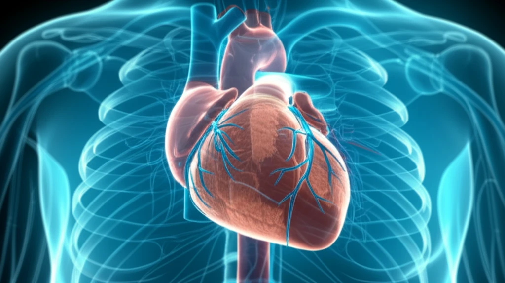
Ventricular tachycardia (VT), a rapid and erratic heartbeat originating in the heart's ventricles, can be life-threatening. Radiofrequency ablation, a common procedure to treat VT, isn't always successful, especially when the source of the arrhythmia lies deep within the heart muscle – a condition known as intramural VT. These deeply seated foci are hard to reach and eliminate with conventional methods, leading to recurrence and the need for repeated interventions.

When standard ablation fails, doctors sometimes turn to transcoronary ethanol ablation (TCEA). This involves injecting ethanol, a type of alcohol, into small coronary arteries that feed the problematic area of the heart, creating a targeted lesion to disrupt the abnormal electrical pathways. However, accurately targeting these arteries is crucial for the procedure's success and to avoid complications.

A recent study published investigates whether using detailed pre-procedural imaging can improve the precision and outcomes of TCEA for refractory septal ventricular tachycardia. The research explores how cardiac magnetic resonance (CMR) and computed tomography (CT) scans can guide the way, offering a potential solution for patients with challenging VT.

Imaging to the Rescue: How Detailed Scans Are Changing the Game

3D heart model showing coronary arteries and scar tissue.

The key innovation explored in this study is the use of advanced imaging techniques before the TCEA procedure. Researchers utilized cardiac magnetic resonance (CMR) and computed tomography (CT) scans to map the heart's structure, identify the precise location of the intramural VT source, and visualize the network of coronary arteries in relation to this source. This detailed roadmap allows doctors to plan the TCEA procedure with much greater accuracy.

The study involved a small group of patients with recurrent septal VT who had already undergone multiple failed radiofrequency ablations. Here's how the pre-procedural imaging was used to guide the TCEA:

  • Mapping the Scar: CMR was used to identify and characterize the scar tissue within the heart muscle, pinpointing the exact location of the arrhythmia's origin.
  • Visualizing the Arteries: CT scans provided a detailed view of the coronary arteries, allowing doctors to select the specific septal perforator branches that supplied blood to the targeted scar tissue.
  • Creating a 3D Roadmap: The CMR and CT images were merged to create a three-dimensional model of the heart, showing the relationship between the scar tissue and the coronary arteries. This roadmap was then integrated into the electroanatomical mapping system used during the TCEA procedure.
After the pre-procedural imaging and careful planning, the TCEA procedure was performed. Ethanol was injected into the selected septal artery branches to create a localized lesion, disrupting the abnormal electrical pathways causing the VT. The researchers then monitored the patients to see how effective the procedure was in reducing their VT burden – the frequency and severity of their arrhythmia.

The Promising Results and Future Directions

The study found that using pre-procedural imaging to guide TCEA led to a significant reduction in VT burden for the patients. The number of ICD therapies (shocks and anti-tachycardia pacing) needed to manage their arrhythmias decreased after the procedure. This suggests that the more targeted approach, thanks to the detailed imaging, can improve the effectiveness of TCEA.

While these results are promising, the researchers emphasize that this is a small, single-center study, so larger investigations are needed to confirm these findings. However, the study provides a strong rationale for using pre-procedural imaging to guide TCEA in carefully selected patients with refractory septal VT.

This approach offers a potential way to improve outcomes for patients with challenging VT, providing a more precise and effective way to target the source of their arrhythmias. As technology advances and imaging techniques become even more sophisticated, we can expect further refinements in this approach, potentially leading to even better outcomes for patients with this difficult condition.

About this Article -

This article was crafted using a human-AI hybrid and collaborative approach. AI assisted our team with initial drafting, research insights, identifying key questions, and image generation. Our human editors guided topic selection, defined the angle, structured the content, ensured factual accuracy and relevance, refined the tone, and conducted thorough editing to deliver helpful, high-quality information.See our About page for more information.

This article is based on research published under:

DOI-LINK: 10.1111/jce.13816, Alternate LINK

Title: Preprocedural Imaging To Guide Transcoronary Ethanol Ablation For Refractory Septal Ventricular Tachycardia

Subject: Physiology (medical)

Journal: Journal of Cardiovascular Electrophysiology

Publisher: Wiley

Authors: Ivo Roca‐Luque, Nuria Rivas‐Gándara, Jaume Francisco‐Pascual, Jose Rodriguez‐Sanchez, Hug Cuellar‐Calabria, Jose Rodriguez‐Palomares, Bruno García‐Del Blanco, Jordi Pérez‐Rodon, Alba Santos‐Ortega, Ferran Rosés‐Noguer, Roger Marsal, Barbara Rubio, David García‐Dorado García, Angel Moya Mitjans

Published: 2019-01-05

Everything You Need To Know

1

What is Ventricular Tachycardia (VT), and why is it a concern?

Ventricular tachycardia (VT) is a rapid and irregular heartbeat originating in the ventricles of the heart. It is a serious condition because it can be life-threatening if left untreated. The article highlights that when standard ablation techniques fail to address VT, especially when the source is deep within the heart muscle (intramural VT), alternative methods like transcoronary ethanol ablation (TCEA) become necessary.

2

What is transcoronary ethanol ablation (TCEA), and why is it used?

Transcoronary ethanol ablation (TCEA) is a procedure used to treat ventricular tachycardia (VT) when standard ablation techniques are unsuccessful. It involves injecting ethanol, a type of alcohol, into small coronary arteries that supply the problematic area of the heart. This creates a targeted lesion to disrupt the abnormal electrical pathways causing the VT. The significance of TCEA lies in its ability to target the source of the arrhythmia directly, especially in cases of intramural VT, where the abnormal pathways are difficult to reach with conventional methods.

3

How do cardiac magnetic resonance (CMR) and computed tomography (CT) scans improve the transcoronary ethanol ablation (TCEA) procedure?

The use of cardiac magnetic resonance (CMR) and computed tomography (CT) scans before a transcoronary ethanol ablation (TCEA) procedure is a key innovation. These imaging techniques provide a detailed map of the heart's structure, pinpointing the exact location of the intramural ventricular tachycardia (VT) source and visualizing the coronary arteries. The importance of these scans lies in their ability to guide the TCEA procedure with greater precision, ensuring that the ethanol is delivered to the correct area. This targeted approach helps to improve the effectiveness of the procedure and reduce complications.

4

How is the pre-procedural imaging used in the process?

The study involved a small group of patients with recurrent septal ventricular tachycardia (VT) who had previously undergone unsuccessful radiofrequency ablations. The article highlights the process that involves using cardiac magnetic resonance (CMR) to map the scar tissue and computed tomography (CT) scans to visualize the coronary arteries. These are then merged to create a 3D roadmap to guide the TCEA procedure. The significance of this process is that it allows doctors to plan the procedure with greater accuracy and to target the abnormal electrical pathways more effectively.

5

What were the key findings of the study regarding the use of pre-procedural imaging for transcoronary ethanol ablation (TCEA)?

The study's results showed a significant reduction in the ventricular tachycardia (VT) burden for patients who underwent transcoronary ethanol ablation (TCEA) guided by pre-procedural imaging. The reduction in the number of ICD therapies (shocks and anti-tachycardia pacing) indicates that the more targeted approach improved the effectiveness of TCEA. This suggests that the use of cardiac magnetic resonance (CMR) and computed tomography (CT) scans to guide the procedure is beneficial for patients with refractory septal VT.

Newsletter Subscribe

Subscribe to get the latest articles and insights directly in your inbox.