Miniature microscope imaging neural networks

Surgical Breakthrough: Minimally Invasive Brain Imaging Revolutionizes Neuroscience

"Discover how a novel technique using skin suturing and surface viral infusion is enhancing neuronal imaging with head-mounted microscopes, offering new insights into brain activity."


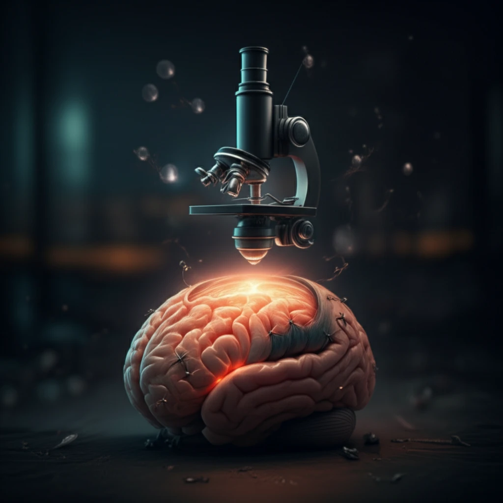
Understanding how the brain works requires observing the activity of large groups of brain cells in real-time. Traditional methods, like two-photon microscopy, often involve securing an animal’s head, which limits natural behaviors. Recent advances have introduced miniature, head-mounted microscopes that allow scientists to study brain activity in freely moving animals.

However, getting clear images through these microscopes is still challenging. Injecting viruses to highlight specific neurons and maintaining clear visibility through cranial windows—surgical openings in the skull—can be tricky. Tissue damage and maintaining window clarity are significant hurdles.

A new approach combines gentle viral infusion techniques with careful skin closure to improve both the labeling of neurons and the long-term clarity of these cranial windows. This method promises to make brain imaging more accessible and reliable.

How Does This New Brain Imaging Technique Work?

Miniature microscope imaging neural networks

The refined surgical technique focuses on two key areas: how neurons are labeled and how the surgical site is managed post-operation. The goal is to minimize damage and maximize the clarity for long-term imaging.

First, instead of directly injecting the virus into the brain tissue, the virus is gently applied to the surface of the cortex using a wide-diameter glass pipette. This helps to label the superficial layers of neurons without causing deep tissue damage. Here's how it breaks down:

  • Surface Viral Infusion: A wide-tip pipette is used to apply the virus to the brain's surface, targeting superficial neurons.
  • Reduced Tissue Damage: This surface application minimizes damage compared to direct injection methods.
  • Minimized Dura Overgrowth: The new method reduces tissue damage and dura overgrowth, which often clouds the window.
After applying the virus and implanting a cranial window, the scalp is carefully sutured back over the surgical site. This protects the window and promotes better healing, reducing inflammation and tissue overgrowth. This is the second critical element in the improved method. Here's why it's important:

The Future of Brain Imaging

By combining surface viral infusion with a sutured scalp technique, researchers are achieving clearer, more reliable brain images over longer periods. This opens new possibilities for studying brain functions in freely moving animals, providing a more natural and accurate understanding of neural processes. The new method not only enhances image quality but also increases the success rate of experiments, allowing for more consistent and reproducible results. This advancement is set to accelerate progress in neuroscience, offering new insights into how the brain works in various behavioral contexts.

About this Article -

This article was crafted using a human-AI hybrid and collaborative approach. AI assisted our team with initial drafting, research insights, identifying key questions, and image generation. Our human editors guided topic selection, defined the angle, structured the content, ensured factual accuracy and relevance, refined the tone, and conducted thorough editing to deliver helpful, high-quality information.See our About page for more information.

Everything You Need To Know

1

What is the primary challenge this new technique addresses in brain imaging?

The primary challenge is achieving clear, long-term brain images, particularly in freely moving animals. Traditional methods often encounter issues with tissue damage, inflammation, and maintaining the clarity of cranial windows, which are necessary for using head-mounted microscopes. This technique aims to overcome these hurdles by refining both neuronal labeling and surgical site management.

2

How does the surface viral infusion method improve neuron labeling compared to traditional techniques?

Instead of direct injection into the brain tissue, the new method uses a wide-diameter glass pipette to gently apply the virus to the cortex's surface. This approach, referred to as Surface Viral Infusion, targets the superficial layers of neurons. The advantage is reduced tissue damage compared to direct injection, leading to better imaging clarity and a higher success rate in studies.

3

Why is careful skin closure, achieved through suturing, so important in this new brain imaging technique?

Careful skin closure, through suturing, is critical because it protects the cranial window and promotes better healing. This reduces inflammation and tissue overgrowth, which often cloud the window and impair imaging quality. By suturing the scalp back over the surgical site, the technique helps maintain long-term clarity and reliability of brain images.

4

What are the key elements of the surgical breakthrough that improves brain imaging?

The surgical breakthrough combines two key elements: Surface Viral Infusion for neuron labeling and the careful suturing of the scalp for surgical site management. The Surface Viral Infusion uses a wide-tip pipette to apply the virus, reducing damage to neurons. Following the viral infusion, careful skin closure using sutures is performed to protect the cranial window and reduce inflammation and tissue overgrowth, which leads to better clarity.

5

How does this new approach impact the study of brain function in freely moving animals and the future of neuroscience?

By combining Surface Viral Infusion with careful skin closure, the new method enables researchers to achieve clearer and more reliable brain images over longer periods. This enhancement is particularly beneficial for studying brain functions in freely moving animals, allowing for a more natural and accurate understanding of neural processes. This technique enhances image quality and increases the success rate of experiments, leading to more consistent and reproducible results, which promises to accelerate advancements in neuroscience.

Newsletter Subscribe

Subscribe to get the latest articles and insights directly in your inbox.