Illustration of a brain highlighting a granular cell tumor near the pituitary gland.

Sudden Vision Changes? What You Need to Know About Granular Cell Tumors

"Understanding a rare brain tumor: Symptoms, diagnosis, and treatment options for granular cell tumors affecting the pituitary gland."


Imagine experiencing a sudden loss of vision, a frightening event that can significantly disrupt your life. While many factors can contribute to vision changes, one rare cause is a granular cell tumor (GCT) affecting the pituitary gland. This article explores this uncommon type of brain tumor, shedding light on its characteristics, diagnosis, and management.

Granular cell tumors are typically benign, slow-growing neoplasms. However, their location near critical brain structures like the optic chiasm and pituitary gland can lead to various neurological symptoms. Understanding these tumors is crucial for early detection and appropriate intervention.

This information is based on a case study of a 57-year-old woman who experienced transient vision loss due to a GCT. By delving into this case and the broader research on GCTs, we aim to provide a comprehensive overview accessible to everyone, regardless of their medical background.

What are Granular Cell Tumors?

Illustration of a brain highlighting a granular cell tumor near the pituitary gland.

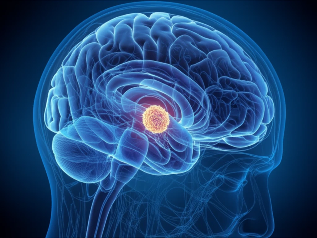
Granular cell tumors (GCTs) are uncommon neoplasms that most often arise in association with the posterior pituitary or infundibulum, a region within the brain. Though often microscopic and discovered during autopsy, some GCTs can grow and cause symptoms.

These tumors are more frequently diagnosed in women between their fourth and fifth decades of life. When symptomatic, the common indications are visual deficits, headaches, hyperprolactinemia, and amenorrhea; less than 5% of those diagnosed present with diabetes insipidus.

  • Visual Disturbances: Compression of the optic chiasm can lead to vision loss.
  • Headaches: Pressure on surrounding brain tissue may trigger headaches.
  • Hormonal Imbalances: Interference with pituitary function can cause hyperprolactinemia (excessive prolactin production) and amenorrhea (absence of menstruation).
  • Diabetes Insipidus: In rare cases, damage to the pituitary stalk can disrupt vasopressin production, leading to diabetes insipidus.
The location of the tumor, its growth pattern, and the way it presents itself clinically all help doctors reach a diagnosis. Microscopic examination is key, revealing distinctive features such as sheets of large polygonal cells containing eccentrically placed nuclei. A granular content is shown in the generous, eosinophilic cytoplasm.

Navigating Diagnosis and Treatment

Diagnosing a granular cell tumor often involves a combination of neuroimaging techniques, such as MRI and CT scans, and a thorough clinical evaluation. MRI can help visualize the tumor's location, size, and relationship to surrounding structures. The lack of expansion of the sella turcica and the presence of normal enhancing pituitary tissue within the sella, but without tumor continuity, limited the initial differential diagnosis.

Treatment typically involves surgical resection to remove the tumor and alleviate symptoms. However, GCTs can be firm and vascular, so a total resection is not always possible. In cases where the tumor cannot be completely removed, adjuvant radiation therapy may be recommended.

If you're experiencing sudden vision changes or other neurological symptoms, prompt medical evaluation is essential. While granular cell tumors are rare, early diagnosis and appropriate treatment can help manage symptoms and improve outcomes. Talk to your doctor to get a diagnosis and personalized treatment plan.

About this Article -

This article was crafted using a human-AI hybrid and collaborative approach. AI assisted our team with initial drafting, research insights, identifying key questions, and image generation. Our human editors guided topic selection, defined the angle, structured the content, ensured factual accuracy and relevance, refined the tone, and conducted thorough editing to deliver helpful, high-quality information.See our About page for more information.

Everything You Need To Know

1

What exactly are Granular Cell Tumors?

Granular cell tumors (GCTs) are rare, slow-growing neoplasms. They are typically found in the neurohypophysis, which includes the posterior pituitary and infundibulum, areas within the brain. These tumors are often microscopic but can grow large enough to cause symptoms. They are considered important because of their potential to cause vision problems and other neurological issues due to their location near critical brain structures such as the optic chiasm and pituitary gland. Implications of GCTs can include visual disturbances, headaches, and hormonal imbalances.

2

What are the typical symptoms of Granular Cell Tumors?

Common symptoms of a granular cell tumor (GCT) include visual disturbances, headaches, hyperprolactinemia, and amenorrhea. Visual disturbances arise from compression of the optic chiasm, potentially leading to vision loss. Headaches result from pressure on surrounding brain tissue. Hormonal imbalances, like hyperprolactinemia and amenorrhea, occur due to the interference with pituitary function. Less than 5% of those diagnosed with GCTs present with diabetes insipidus, which is caused by the damage to the pituitary stalk disrupting vasopressin production.

3

How are Granular Cell Tumors usually diagnosed?

Diagnosing a granular cell tumor (GCT) involves several steps. Neuroimaging techniques, such as MRI and CT scans, are used to visualize the tumor's location, size, and its relationship with the surrounding structures. The lack of expansion of the sella turcica and the presence of normal enhancing pituitary tissue are key indicators. Microscopic examination is essential, revealing distinctive features such as sheets of large polygonal cells containing eccentrically placed nuclei and a granular content within the cytoplasm. These diagnostic steps help doctors to understand the location and presentation of the tumor, leading to the diagnosis.

4

How can a Granular Cell Tumor cause vision problems?

Granular cell tumors (GCTs) can lead to vision problems due to their location near critical brain structures, particularly the optic chiasm. The optic chiasm is where the optic nerves from each eye cross. As a GCT grows, it can compress this structure. This compression can result in visual deficits and vision loss. Understanding this relationship is crucial for early detection and appropriate intervention to prevent further vision deterioration.

5

How are Granular Cell Tumors usually treated?

Treatment for granular cell tumors (GCTs) is not explicitly detailed but is implied to include a combination of diagnosis and management strategies. The provided information highlights the importance of early detection and appropriate intervention. The diagnosis relies on a combination of neuroimaging techniques, like MRI and CT scans, and a thorough clinical evaluation. Management would likely involve monitoring the tumor, medication to manage symptoms, or potential surgical intervention, depending on the tumor's size, location, and impact on the patient's health. The goal is to address the neurological symptoms, visual disturbances, and hormonal imbalances associated with the tumor.

Newsletter Subscribe

Subscribe to get the latest articles and insights directly in your inbox.