Surreal endoscopic view of digestive tract tumors.

Stomach Submucosal Tumors: A Comprehensive Guide

"From detection to treatment, understand the landscape of stomach submucosal tumors and their management."


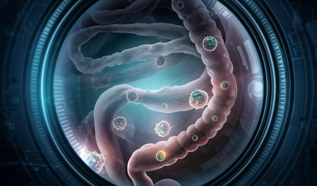
Stomach submucosal tumors (SMTs) encompass a variety of lesions within the digestive tract. These lesions develop from different layers of the digestive tube, ranging from deep within the mucosa to the serosa, and may originate from tumors or other non-tumorous sources. While the term 'subepithelial lesions' might be more accurate, SMT remains the preferred term in common practice, especially in endoscopy.

Distinguishing these lesions is crucial because some carry a potential for malignancy. SMTs are detected in approximately one out of every 300 endoscopies, with about 60% located in the stomach. Gastric SMTs are often asymptomatic, covered by normal mucosa, and discovered incidentally during endoscopies or radiological examinations. In other instances, digestive bleeding is the most frequent symptom.

When a submucosal-appearing lesion is detected during endoscopy, the initial step involves differentiating it from external compression. Endoscopy alone has limited efficacy for this purpose (39-69%). Endoscopic ultrasound (EUS) is the key examination, superior to endoscopy, transabdominal ultrasound, or computed tomography. EUS demonstrates a sensitivity of 92% and specificity of 100% in differentiating between SMTs and external compression in a prospective study. The subsequent step consists of determining the nature of the SMT, if possible.

Understanding the Nature and Origin of Stomach Tumors

Surreal endoscopic view of digestive tract tumors.

Gastrointestinal stromal tumors (GISTs) are the most common mesenchymal tumors of the digestive tract, developing at the expense of Cajal cells or one of their precursors. They reside in approximately 65% of cases in the stomach and 25% of cases in the small intestine. GISTs are now perfectly defined on the nosological level, in particular by the expression in immunohistochemistry of the KIT protein (95% of cases). An activating mutation of the KIT genes or more rarely PDGFRA is found in approximately 85% of cases. Their potential for malignancy is all the more important as their size and their mitotic index are high.

The leimyomas are benign spindle cell tumors developed from the smooth muscle cells of the muscularis mucosae or the muscularis propria. The best knowledge of GIST has shown that the leiomyomas are rare in the stomach, but are mostly located in the esophagus, and less frequently in the rectum. The histological distinction between GIST and leimyomas is mainly based on immunohistochemistry, the leimyomas expressing smooth muscle markers such as actin and desmin, but not the KIT protein.

  • Schwannomas are very rare digestive tumors of nervous origin, benign, whose digestive location most frequently is the proximal part of the stomach. Schwannomas express protein S-100 in immunohistochemistry, but neither KIT nor desmin.
  • Lipoma is a benign tumor consisting of mature fat cells. It is rarer in the stomach than in the colon.
  • Other gastric mesenchymal tumors are exceptional. There are a small number of GISTs that do not express KIT in immunohistochemistry (5%). It is therefore recommended, in these cases where the histology is evocative of GIST, to search for a mutation of the KIT or PDGFRA genes.
The main submucosal lesion of the stomach apart from GIST is not tumoral, since it is an aberrant pancreas. Gastric submucosal non-mesenchymal tumors are essentially constituted by endocrine tumors. Most gastric endocrine tumors are well-differentiated fundus tumors, non-secreting and developed from ECL cells. Three types are distinguished: type I, associated with chronic autoimmune gastritis; type II, associated with multiple neuroendocrine disease (MEN) type 1 and Zollinger-Ellison syndrome; type III, sporadic without hypergastrinemia or chronic autoimmune gastritis.

The Importance of Ongoing Research

The incidental discovery of a gastric SMT during an endoscopy is quite frequent. Recent data has shown that GISTs are the most frequent tumorous lesion. EUS is the key examination for the study of gastric SMTs. It may allow in some cases to establish a presumptive diagnosis and guides the management modalities (monitoring, endoscopic or surgical resection, EUS-guided puncture). Prospective studies are needed to better understand the natural history of SMTs and to better define the role of EUS in the follow-up when a resection is not performed. This would allow to establish recommendations in the management of gastric SMTs that currently exist only for GISTs.

About this Article -

This article was crafted using a human-AI hybrid and collaborative approach. AI assisted our team with initial drafting, research insights, identifying key questions, and image generation. Our human editors guided topic selection, defined the angle, structured the content, ensured factual accuracy and relevance, refined the tone, and conducted thorough editing to deliver helpful, high-quality information.See our About page for more information.

This article is based on research published under:

DOI-LINK: 10.1007/978-2-287-99164-6_16, Alternate LINK

Title: Tumeurs Sous-Muqueuses De L’Estomac

Journal: Écho-endoscopie digestive

Publisher: Springer Paris

Authors: B. Landi, L. Palazzo

Published: 2012-01-01

Everything You Need To Know

1

What exactly are stomach submucosal tumors (SMTs), and why is it important to distinguish between them?

Stomach submucosal tumors, or SMTs, are lesions found in the digestive tract, originating from various layers, including the mucosa and serosa. While 'subepithelial lesions' might be more precise, SMT is the commonly used term, especially in endoscopy. Differentiating these lesions is vital due to the potential for malignancy in some cases. These are detected in approximately one out of every 300 endoscopies, with about 60% located in the stomach.

2

Why is endoscopic ultrasound (EUS) considered a key examination for gastric submucosal tumors, and how does it guide the management of these tumors?

Endoscopic ultrasound, or EUS, is crucial for examining gastric submucosal tumors. EUS demonstrates a sensitivity of 92% and specificity of 100% in differentiating between SMTs and external compression. It can help establish a presumptive diagnosis and guide management, such as monitoring, endoscopic or surgical resection, or EUS-guided puncture. Endoscopy alone has limited efficacy for this purpose (39-69%).

3

What are gastrointestinal stromal tumors (GISTs), and what genetic factors are commonly associated with their development?

Gastrointestinal stromal tumors, or GISTs, are the most common mesenchymal tumors in the digestive tract. They originate from Cajal cells or their precursors, with about 65% found in the stomach and 25% in the small intestine. GISTs are defined by the expression of the KIT protein in immunohistochemistry (95% of cases). An activating mutation of the KIT genes or more rarely PDGFRA is found in approximately 85% of cases.

4

Can you elaborate on leiomyomas and how they are distinguished from gastrointestinal stromal tumors (GISTs) in the stomach?

Leiomyomas are benign spindle cell tumors arising from the smooth muscle cells of the muscularis mucosae or muscularis propria. They are rare in the stomach, mostly located in the esophagus, and less frequently in the rectum. Histological distinction between GIST and leiomyomas is based on immunohistochemistry, where leiomyomas express smooth muscle markers like actin and desmin, but not the KIT protein.

5

Besides GISTs, what other types of tumors and non-tumorous lesions can manifest as submucosal masses in the stomach, particularly concerning endocrine tumors?

Gastric submucosal non-mesenchymal tumors primarily consist of endocrine tumors. Most gastric endocrine tumors are well-differentiated fundus tumors, non-secreting, and developed from ECL cells. Three types exist: type I, linked to chronic autoimmune gastritis; type II, linked to multiple neuroendocrine disease (MEN) type 1 and Zollinger-Ellison syndrome; type III, sporadic without hypergastrinemia or chronic autoimmune gastritis. The main submucosal lesion of the stomach apart from GIST is an aberrant pancreas.

Newsletter Subscribe

Subscribe to get the latest articles and insights directly in your inbox.