AI-enhanced rib fracture detection.

Spotting Rib Fractures: How Tech Can Help Doctors See the Unseen

"AI-powered tools are stepping in to help doctors quickly and accurately diagnose rib fractures, potentially improving patient outcomes and reducing missed injuries."


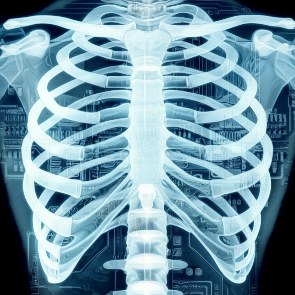
Rib fractures are a common injury, often resulting from falls, car accidents, or sports-related trauma. Detecting these fractures accurately is crucial for managing pain, preventing complications like pneumonia, and identifying underlying issues. However, spotting rib fractures on chest X-rays can be challenging, even for experienced doctors.

Traditional methods of detection heavily rely on the radiologist's expertise and the quality of the X-ray image. This can lead to missed fractures or delayed diagnoses, especially since ribs are small, overlap each other, and can be obscured by other structures in the chest. Up to 50% of rib fractures can be missed with radiography alone, putting patients at risk for further complications.

Now, researchers are exploring new technologies to aid in rib fracture detection. One promising approach involves using computer-aided methods to analyze chest X-rays. These methods use local texture and shape features to quantify the characteristics of broken ribs. By using these measurements, medical professionals can accurately identify the fractures for quick and effective treatment.

AI to the Rescue: How the Measurement Method Works

AI-enhanced rib fracture detection.

Researchers have developed a new measurement method to assist doctors in identifying rib fractures on chest X-rays. This method uses a two-step process:

First, the system assigns a measurement area around each rib using a spline curve. This creates a customized outline that follows the unique shape of each rib. Sampling lines are then generated perpendicular to the spline curve, allowing for detailed analysis of the rib's structure.

  • Measurement Area Assignment: Uses a spline curve to adapt to the rib's shape.
  • Fracture-ness Measurement: Applies asymmetry and texture analysis.
  • ROI Analysis: Identifies fracture and normal regions.
  • SVM Classification: Uses machine learning to classify fractures.
Second, the system analyzes the 'fracture-ness' of the rib using three key measurements: asymmetry, contrast, and homogeneity. Asymmetry measures how similar the two sides of the rib are, contrast highlights differences in pixel intensity, and homogeneity assesses the uniformity of the rib's texture. These measurements help to identify deviations from a normal rib structure, indicating a possible fracture.

The Future of Fracture Detection

This new measurement method shows great promise in improving the accuracy and speed of rib fracture detection. In tests, the method was able to identify statistically significant differences between fractured and normal ribs.

While this technology is not meant to replace radiologists, it can serve as a valuable tool to aid in diagnosis and potentially reduce the number of missed fractures. By providing doctors with a more detailed and objective analysis of chest X-rays, computer-aided detection methods can lead to better patient outcomes.

Further research is needed to refine these techniques and test them on larger patient populations. However, the initial results are encouraging and suggest that AI-powered tools could play a significant role in the future of fracture detection.

About this Article -

This article was crafted using a human-AI hybrid and collaborative approach. AI assisted our team with initial drafting, research insights, identifying key questions, and image generation. Our human editors guided topic selection, defined the angle, structured the content, ensured factual accuracy and relevance, refined the tone, and conducted thorough editing to deliver helpful, high-quality information.See our About page for more information.

This article is based on research published under:

DOI-LINK: 10.4258/hir.2013.19.3.196, Alternate LINK

Title: Quantitative Measurement Method For Possible Rib Fractures In Chest Radiographs

Subject: Health Information Management

Journal: Healthcare Informatics Research

Publisher: The Korean Society of Medical Informatics

Authors: Jaeil Kim, Sungjun Kim, Young Jae Kim, Kwang Gi Kim, Jinah Park

Published: 2013-01-01

Everything You Need To Know

1

What makes spotting rib fractures on chest X-rays so difficult?

Rib fractures are often caused by falls, car accidents, or sports injuries. The main challenge with detecting them is that ribs are small and can be hidden by other structures on chest X-rays. Traditional methods rely on a radiologist's expertise and the quality of the image, which can lead to missed fractures or delayed diagnoses. Up to 50% of rib fractures can be missed using just radiography. These missed or delayed diagnoses can lead to further health issues.

2

How does the new measurement method work?

The new measurement method uses a two-step process to identify rib fractures. First, it assigns a measurement area around each rib using a spline curve. This creates a customized outline. Next, the system analyzes the fracture-ness of the rib using three key measurements: asymmetry, contrast, and homogeneity. Asymmetry measures the similarity between the two sides of the rib. Contrast highlights differences in pixel intensity. Homogeneity assesses the uniformity of the rib's texture. These measurements help identify deviations from a normal rib structure, indicating a possible fracture. After this the method applies region of interest (ROI) analysis and support vector machine (SVM) classification.

3

What measurements are used to determine the 'fracture-ness' of a rib?

The 'fracture-ness' is determined by assessing asymmetry, contrast, and homogeneity. Asymmetry checks the similarity of the rib's two sides; contrast emphasizes differences in pixel intensity, showing variations in the bone structure; and homogeneity evaluates the uniformity of the rib's texture. These three measurements help to pinpoint areas that deviate from the normal structure, potentially indicating a fracture. The AI uses these features, combined with the data from the region of interest (ROI) analysis and the support vector machine (SVM) classification to classify and identify the fracture.

4

How does the spline curve contribute to rib fracture detection?

The spline curve is used in the measurement area assignment step. The system uses the spline curve to create a custom outline that follows each rib's unique shape. This allows the system to analyze the rib's structure in detail. Sampling lines are generated perpendicular to the spline curve, allowing for detailed analysis of the rib's structure.

5

Why is using AI for rib fracture detection important?

AI-powered tools are significant because they can improve the accuracy and speed of rib fracture detection. They offer a more reliable method than traditional approaches that heavily rely on a radiologist's skill. This could lead to earlier diagnoses and quicker treatment for patients. Earlier diagnosis can prevent complications like pneumonia and help to identify underlying issues. The results showed the method was able to identify statistically significant differences between fractured and normal ribs.

Newsletter Subscribe

Subscribe to get the latest articles and insights directly in your inbox.