Illustration of spinal cord regeneration using a biodegradable implant.

Spinal Cord Breakthrough: Can FGF1 and Nerve Grafts Restore Movement?

"New research explores how a biodegradable device delivering FGF1 and nerve grafts could revolutionize spinal cord injury treatment and restore motor function."


Spinal cord injury (SCI) presents a formidable challenge in neuroscience. While significant progress has been made in recent years, finding effective strategies to repair and restore function after SCI remains a primary goal. Current research focuses on several key areas, including limiting secondary damage, preventing further neuronal damage, promoting regeneration across the injury site, and improving the local environment to support healing.

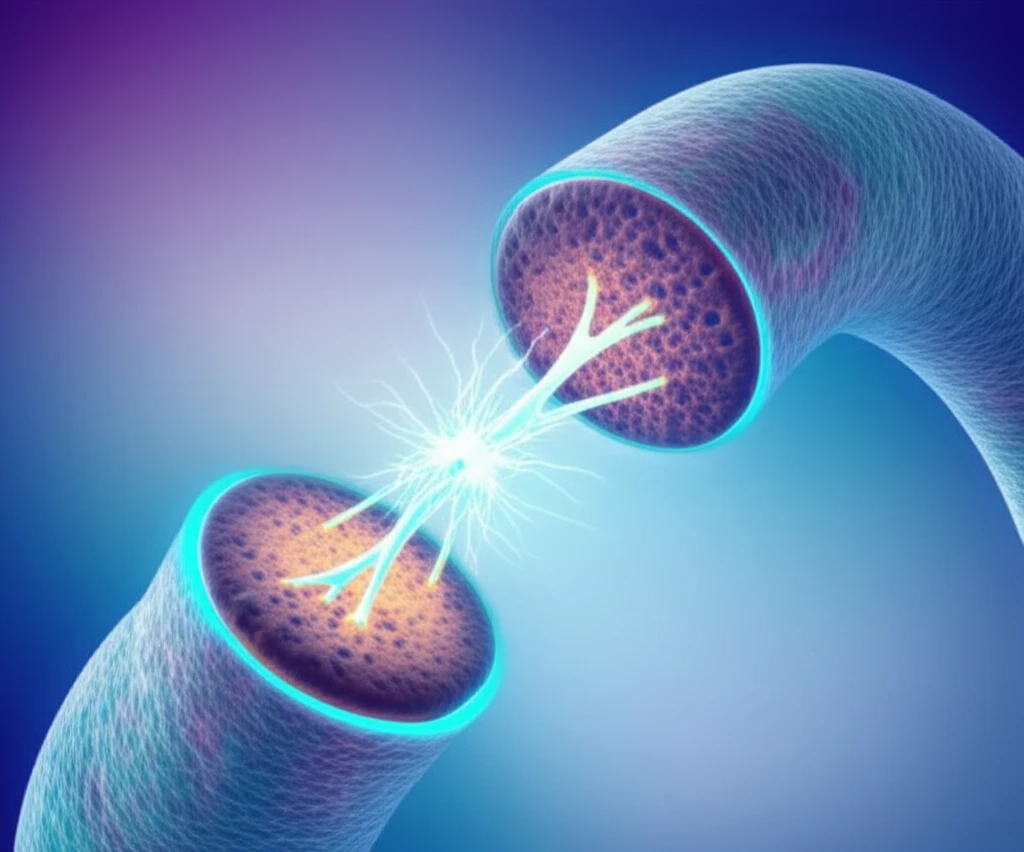
One promising avenue involves the use of peripheral nerve grafts (PNGs). These grafts can act as a bridge, guiding regenerating nerve fibers across the damaged area of the spinal cord. Some approaches combine PNGs with acidic fibroblast growth factor (FGF1) to enhance nerve regeneration and redirect nerve fibers into adjacent gray matter, potentially bypassing inhibitory factors in the white matter.

Recognizing the need for precision in PNG placement, researchers have developed a graft holder device to improve surgical accuracy. In pursuit of a translational development, this article looks into a new biodegradable version that may be absorbed by the body, preventing long-term complications associated with implanted materials. This new device will contain PNGs, with or without FGF1.

Can a New Device with FGF1 and Nerve Grafts Improve Spinal Cord Repair?

Illustration of spinal cord regeneration using a biodegradable implant.

The study published in Restorative Neurology and Neuroscience explores a novel approach to spinal cord repair in rats using a biodegradable device containing peripheral nerve grafts (PNGs) and fibroblast growth factor 1 (FGF1). The aim was to assess whether this combination could promote nerve regeneration, restore motor function, and integrate effectively with the spinal cord tissue.

Researchers subjected rats to a T11 spinal cord resection and subsequently implanted twelve PNGs oriented towards the white-to-grey matter within a biodegradable device, with or without FGF1. The success of the treatments were tracked by...

  • BBB Score: To quantify hindlimb motor function recovery.
  • Electrophysiology: To measure motor evoked potentials (MEPs), indicating functional connections between the brain and muscles.
  • Immunohistochemistry: To examine nerve fiber regeneration and the presence of corticospinal tract fibers using anterograde BDA tracing.
The results revealed promising outcomes. Motor evoked potentials (MEPs) reappeared in the lower limbs of treated animals at 20 weeks post-grafting, with significant improvements observed in the group receiving FGF1. This reappearance of MEPs was paralleled by the presence of NF-positive fibers and anterogradely traced corticospinal fibers distal to the injury site, indicating successful nerve regeneration. Furthermore, the BBB scores improved in the treated animals, suggesting enhanced motor function.

Future of Spinal Cord Regeneration

These findings support the potential of combining PNGs and FGF1 as a strategy for spinal cord reinnervation. The innovative biodegradable device facilitated robust MEPs, augmented by FGF1, marking a significant step towards translational research and potential clinical applications. Further studies are needed to optimize this approach and assess its long-term efficacy, but the initial results offer hope for improved outcomes in spinal cord injury treatment.

About this Article -

This article was crafted using a human-AI hybrid and collaborative approach. AI assisted our team with initial drafting, research insights, identifying key questions, and image generation. Our human editors guided topic selection, defined the angle, structured the content, ensured factual accuracy and relevance, refined the tone, and conducted thorough editing to deliver helpful, high-quality information.See our About page for more information.

Everything You Need To Know

1

What is the primary goal of the research described and what is the main approach?

The primary goal is to find effective strategies to repair and restore function after Spinal Cord Injury (SCI). The main approach involves the use of a biodegradable device containing peripheral nerve grafts (PNGs) and fibroblast growth factor 1 (FGF1) to promote nerve regeneration and restore motor function. This combination aims to bridge the damaged area of the spinal cord, enhance nerve regeneration, and potentially bypass inhibitory factors, ultimately improving the local environment to support healing.

2

How do peripheral nerve grafts (PNGs) and FGF1 contribute to spinal cord repair?

Peripheral nerve grafts (PNGs) act as a bridge, guiding regenerating nerve fibers across the damaged area of the spinal cord. FGF1 enhances nerve regeneration and redirects nerve fibers into adjacent gray matter, potentially bypassing inhibitory factors present in the white matter. The combination of PNGs and FGF1 within a biodegradable device is designed to create an environment conducive to nerve fiber regrowth and functional recovery.

3

What is the significance of the biodegradable device in this research and why is it important?

The biodegradable device is designed to contain PNGs, with or without FGF1, and it is absorbed by the body, preventing long-term complications associated with implanted materials. The device is crucial for precise placement of the grafts and for improving surgical accuracy. The use of a biodegradable device represents a step towards translational development and potential clinical applications, as it minimizes the need for future interventions related to the device itself.

4

What specific methods were used to assess the success of the spinal cord repair treatment, and what did the results show?

The success of the spinal cord repair treatment was assessed using the BBB Score to quantify hindlimb motor function recovery, Electrophysiology to measure motor evoked potentials (MEPs), and Immunohistochemistry to examine nerve fiber regeneration. The results revealed that MEPs reappeared in the lower limbs of treated animals, with significant improvements in the group receiving FGF1. The BBB scores improved, and nerve fiber regeneration was observed, indicating enhanced motor function and successful nerve regeneration distal to the injury site.

5

What are the future implications of this research, and what further steps are needed?

This research supports the potential of combining PNGs and FGF1 as a strategy for spinal cord reinnervation. The biodegradable device facilitated robust MEPs, augmented by FGF1, indicating a significant step towards translational research and potential clinical applications. Further studies are needed to optimize this approach and assess its long-term efficacy, including detailed assessments of the long-term effects and potential side effects, and scaling up the research to larger animal models before human clinical trials.

Newsletter Subscribe

Subscribe to get the latest articles and insights directly in your inbox.