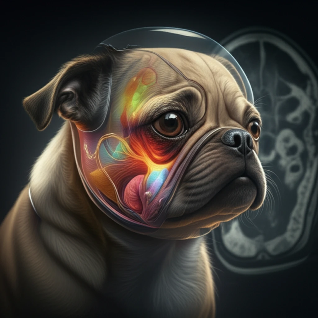
MRI reveals airway anatomy of brachycephalic dog

Snoring and Short Snouts: How MRI is Changing the Game for Brachycephalic Dogs

"Advanced imaging reveals the hidden airway issues in breeds like Pugs and Bulldogs, paving the way for better treatments and happier pets."


If you're a pet parent to a Bulldog, Pug, or Shih Tzu, you're likely familiar with the snorts, snores, and sometimes, the outright struggles to breathe that come with these breeds. Known as brachycephalic dogs, these companions have been selectively bred for their adorable, flat faces. However, this charming trait comes with a significant downside: brachycephalic obstructive airway syndrome (BOAS).

BOAS is a condition that affects the upper airways, making it difficult for these dogs to get enough air. The shortened facial structure leads to a crowded airway, often involving stenotic nares (narrowed nostrils), elongated soft palates, and tracheal hypoplasia (a smaller windpipe). Imagine trying to breathe through a pinched straw—that's the daily reality for many brachycephalic dogs.

Traditionally, diagnosing and understanding BOAS has been challenging, relying on physical exams and sometimes invasive procedures. But now, advancements in veterinary imaging, particularly magnetic resonance imaging (MRI), are providing a clearer picture of what's happening inside these dogs. A recent study published in 'The Journal of Veterinary Medical Science' explores how MRI is revolutionizing our understanding and treatment of BOAS, offering hope for improving the lives of our beloved flat-faced friends.

MRI: A Window into the Brachycephalic Airway

MRI reveals airway anatomy of brachycephalic dog

Magnetic Resonance Imaging (MRI) is non-invasive and provides a superior resolution to view the soft tissues composing the upper airway. It’s accurate, reproducible, and doesn’t use ionizing radiation, making it a safe option for repeated evaluations. Unlike X-rays or CT scans, MRI excels at differentiating between different types of soft tissues. This is crucial when assessing the complex anatomy of a brachycephalic dog's airway, where subtle changes can significantly impact breathing.

The study from 'The Journal of Veterinary Medical Science' utilized MRI to compare the airways of brachycephalic and non-brachycephalic dogs, focusing on the soft palate and nasopharynx, and here is what the MRI revealed:

  • Significant Differences: The MRI scans revealed notable differences in skull length, soft palate length-to-skull ratio, and soft palate thickness between brachycephalic and non-brachycephalic breeds.
  • Volume Matters: Brachycephalic dogs had smaller nasopharyngeal volumes, meaning less space for air to pass through.
  • Soft Palate Shape: The shape of the soft palate varied significantly. Brachycephalic dogs often had thicker, more irregular soft palates that further obstructed their airways.
These findings underscore that it's not just about having a shorter face; the specific dimensions and shapes of the soft tissues within the airway play a critical role in BOAS. MRI allows vets to pinpoint these issues with unprecedented accuracy.

Looking Ahead: Better Breathing for Brachycephalic Breeds

The use of MRI in assessing brachycephalic dogs is more than just a diagnostic tool; it's a pathway to better, more targeted treatments. By understanding the specific anatomical challenges each dog faces, veterinarians can tailor surgical interventions or management strategies to maximize their effectiveness. As research continues, MRI will likely play an increasingly important role in helping brachycephalic breeds breathe easier and live happier, healthier lives.

About this Article -

This article was crafted using a human-AI hybrid and collaborative approach. AI assisted our team with initial drafting, research insights, identifying key questions, and image generation. Our human editors guided topic selection, defined the angle, structured the content, ensured factual accuracy and relevance, refined the tone, and conducted thorough editing to deliver helpful, high-quality information.See our About page for more information.

Everything You Need To Know

1

What is Brachycephalic Obstructive Airway Syndrome (BOAS), and why is it a concern?

Brachycephalic Obstructive Airway Syndrome (BOAS) is a condition prevalent in breeds with shortened faces, like Bulldogs and Pugs. It arises from the unique anatomical challenges associated with their compressed facial structure, leading to obstructed upper airways. The significance of BOAS lies in its impact on a dog's ability to breathe properly, affecting their overall health and quality of life. Implications range from exercise intolerance and sleep disturbances to severe respiratory distress, highlighting the need for accurate diagnosis and tailored management strategies to alleviate suffering and improve the well-being of affected dogs.

2

What is an MRI, and why is it helpful for understanding breathing problems in certain dog breeds?

Magnetic Resonance Imaging (MRI) is a non-invasive imaging technique that provides detailed views of the soft tissues in a dog's airway. It is important because it allows veterinarians to accurately assess the specific anatomical abnormalities contributing to Brachycephalic Obstructive Airway Syndrome (BOAS). The implications of using MRI include better diagnosis, more targeted treatments, and improved outcomes for dogs with BOAS, as it helps identify issues like stenotic nares, elongated soft palates, and tracheal hypoplasia without the need for invasive procedures. Furthermore, MRI's ability to differentiate between various soft tissues offers a distinct advantage over other imaging methods like X-rays or CT scans.

3

What is the soft palate, and how can it cause breathing issues in brachycephalic dogs?

The soft palate is a flap of tissue located at the back of the throat. In brachycephalic dogs, the soft palate is often elongated or thickened, contributing to airway obstruction. The significance of soft palate abnormalities is that they directly impede airflow, exacerbating breathing difficulties in affected dogs. The implications include increased snoring, difficulty exercising, and potential for respiratory distress, underscoring the importance of assessing and, if necessary, surgically correcting soft palate issues to improve breathing.

4

What are stenotic nares, and how do they affect breathing?

Stenotic nares are narrowed nostrils that restrict airflow into the respiratory system. These are significant because they represent a primary point of obstruction, making it harder for brachycephalic dogs to inhale. The implications of stenotic nares include increased effort to breathe, reduced oxygen intake, and exacerbation of other Brachycephalic Obstructive Airway Syndrome (BOAS)-related issues. Surgical correction of stenotic nares is often recommended to improve airflow and alleviate respiratory distress.

5

What is tracheal hypoplasia, and what are the implications for dogs affected by it?

Tracheal hypoplasia refers to an abnormally small trachea (windpipe). It is important because it restricts the amount of air that can flow into the lungs. The implications of tracheal hypoplasia are significant, as it can lead to chronic respiratory problems, reduced exercise tolerance, and increased risk during anesthesia or periods of stress. While surgical correction is not always feasible, understanding the presence and severity of tracheal hypoplasia is crucial for managing a brachycephalic dog's respiratory health and minimizing potential complications. Management strategies may include weight management, avoiding strenuous exercise, and careful monitoring during veterinary procedures.

Newsletter Subscribe

Subscribe to get the latest articles and insights directly in your inbox.