Skin Deep: How Motion Correction Technology is Revolutionizing Dermatology
"Discover how new motion correction algorithms are enhancing optoacoustic mesoscopy, providing clearer and more reliable skin imaging for better diagnoses and treatment."
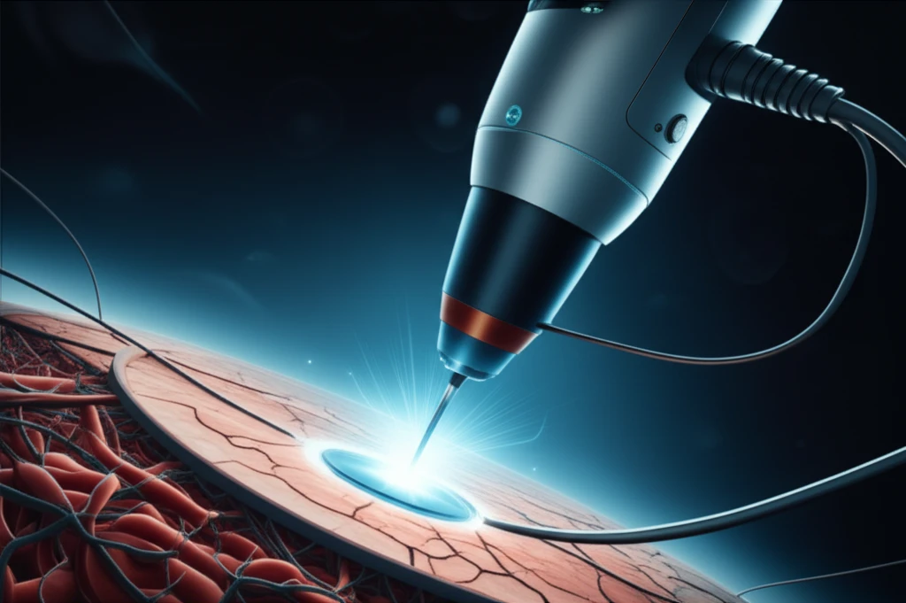
In the ever-evolving field of dermatology, the quest for clearer, more accurate imaging techniques is ongoing. Traditional methods often fall short when dealing with the complexities of skin analysis, particularly at deeper levels. However, a groundbreaking technology known as optoacoustic mesoscopy (RSOM), or photoacoustic mesoscopy, is changing the landscape. This innovative approach provides unprecedented insights into vascular morphology and the pathophysiological biomarkers of skin inflammation, reaching depths that other optical methods simply cannot.
RSOM utilizes ultra-wideband detection and focused ultrasound transducers to achieve remarkable axial resolution of 4 microns and lateral resolution of 20 microns, penetrating several millimeters beneath the skin's surface. While this technology holds immense promise, it faces a significant challenge: motion. Movements, whether from breathing or slight shifts in position, can severely compromise image quality and reduce the effective resolution.
To address this critical issue, researchers have developed a sophisticated motion correction algorithm designed specifically for RSOM. This algorithm analyzes disruptions in the ultrasound wave front caused by the vertical movement of the melanin layer at the skin surface. By generating a smooth, synthetic surface from these disruptions, the algorithm can correct the relative position of the ultrasound detector, leading to dramatically improved image clarity and diagnostic accuracy.
How Motion Correction Works
The motion correction algorithm represents a significant leap forward in medical imaging. It works by meticulously tracking the skin surface, identifying distortions caused by movement during the scanning process. These distortions are primarily observed in the ultrasound wave front generated by melanin-containing layers within the skin.
- Detection of the Disrupted Skin Surface: The algorithm first identifies discontinuities in the skin surface within the three-dimensional sinogram, using different approaches based on the type of skin being examined. For hairless skin, a two-dimensional parabolic slab is used, while for hairy skin, a low-frequency footprint is exploited.
- Generation of an Artificial Continuous Surface: Once the disrupted surface is detected, the algorithm generates a smooth, continuous surface using a moving average filter. The difference between the actual and artificial surfaces indicates the vertical movement of the skin, which is then used to adjust the focal point of the detector during image reconstruction.
The Future of Dermatology
With the ability to correct for motion artifacts and achieve unprecedented resolution, RSOM is poised to become an indispensable tool for dermatologists. Whether it's assessing angiogenesis in melanoma tumors, quantifying inflammation biomarkers, or measuring blood oxygenation and melanin content, this technology offers a wealth of information that was previously unattainable. As RSOM continues to evolve, its impact on dermatology will only grow, leading to earlier diagnoses, more effective treatments, and ultimately, better outcomes for patients.