Advanced medical scanner projecting a clear, detailed image.

Shorter Scans, Clearer Images: How New Imaging Tech is Changing Healthcare

"A breakthrough in combined kV/MV CBCT imaging uses a high-DQE MV detector to improve image quality and reduce scan times, potentially revolutionizing radiotherapy and diagnostic imaging."


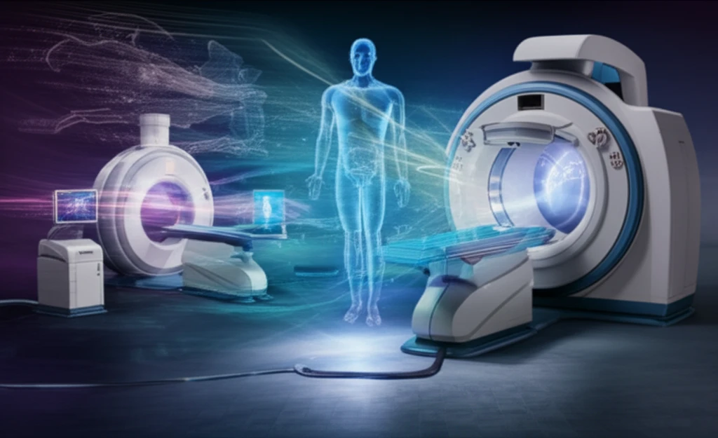
In the rapidly evolving world of medical imaging, precision and speed are paramount. State-of-the-art medical linear accelerators now commonly integrate two imaging systems: an electronic portal imaging device (EPID) used with the treatment beam, and an orthogonal kilovoltage (kV) system for high sensitivity. These systems offer complementary information. The combination of these technologies is paving the way for more accurate diagnoses and more effective treatments.

Traditionally, kV cone-beam tomography (CBCT) and kV planar imaging have been essential for patient setup, beam gating, and delivery verification, enhancing the localization accuracy for various medical procedures. While MV (megavoltage) systems are typically used for treatment quality assurance and exit dosimetry, advancements are making them increasingly relevant for patient setup and beam gating as well.

Recent research suggests that combining kV and MV imaging could revolutionize medical imaging. This innovative approach can significantly reduce scan times and minimize metal artifacts, leading to clearer and more detailed images. By merging orthogonal kV and MV projections, acquired during a short gantry rotation, healthcare professionals can obtain comprehensive data more efficiently.

What is Combined kV/MV CBCT Imaging and Why Does It Matter?

Advanced medical scanner projecting a clear, detailed image.

Combined kV/MV CBCT imaging is an advanced technique that merges data from both kilovoltage (kV) and megavoltage (MV) imaging systems. This innovative approach addresses limitations of traditional imaging methods and has the potential for significant improvements in image-guided radiotherapy and diagnostic imaging.

The primary goals of combined kV/MV CBCT imaging are to:

  • Reduce Scan Time: By combining data from different imaging sources, the total time required to complete a scan can be drastically reduced.
  • Minimize Metal Artifacts: Metal implants and other foreign objects can cause distortions in medical images. Combined imaging techniques help to correct these distortions, providing clearer images.
However, the feasibility of combined kV/MV CBCT imaging has been questioned due to the low detective quantum efficiencies (DQEs) of commercially available electronic portal imagers (EPIDs). The key is developing and testing high DQE MV detectors that can produce acceptable quality pre-treatment CBCT images at acceptable dose levels. A new study provides insight into how this can be achieved.

The Future of Medical Imaging is Here

The integration of high-DQE MV detectors into combined kV/MV imaging systems represents a significant leap forward in medical imaging technology. By reducing scan times and minimizing metal artifacts, this approach promises to enhance diagnostic accuracy, improve treatment outcomes, and provide a better overall experience for patients. As research continues and technology advances, we can anticipate even more groundbreaking developments that will shape the future of healthcare.

About this Article -

This article was crafted using a human-AI hybrid and collaborative approach. AI assisted our team with initial drafting, research insights, identifying key questions, and image generation. Our human editors guided topic selection, defined the angle, structured the content, ensured factual accuracy and relevance, refined the tone, and conducted thorough editing to deliver helpful, high-quality information.See our About page for more information.

Everything You Need To Know

1

What is the core innovation in this new imaging technology?

The core innovation lies in combined kV/MV CBCT imaging. This technique merges data from kilovoltage (kV) and megavoltage (MV) imaging systems to overcome limitations of traditional methods. This combination aims to improve image quality, reduce scan times, and enhance diagnostic accuracy in radiotherapy and diagnostic imaging.

2

How does combined kV/MV CBCT imaging improve medical imaging compared to older methods?

Combined kV/MV CBCT imaging offers several key improvements. First, it reduces scan times by efficiently merging data from kV and MV systems. Second, it minimizes metal artifacts, which often distort images in traditional methods. This leads to clearer, more detailed images, improving diagnostic precision and treatment planning, especially in situations involving metal implants or foreign objects.

3

What are the roles of kV and MV systems in this new imaging technique?

In combined kV/MV CBCT imaging, the kilovoltage (kV) system and megavoltage (MV) system play complementary roles. The kV system, often orthogonal, provides high sensitivity for patient setup and beam gating, and is used for high-resolution imaging. The MV system, traditionally used for treatment quality assurance, becomes increasingly important for patient setup and beam gating due to advancements in detector technology, contributing to comprehensive data acquisition during short gantry rotations.

4

Why has the use of MV imaging been limited in the past, and what has changed?

Historically, the use of MV imaging was limited by the low detective quantum efficiencies (DQEs) of commercially available electronic portal imagers (EPIDs). These low DQEs resulted in poor image quality. However, new research and technology focus on developing and testing high-DQE MV detectors. With high-DQE MV detectors, acceptable quality pre-treatment CBCT images can be produced at acceptable dose levels, opening up new possibilities in imaging.

5

How does this new imaging technology affect patient care and the future of medical treatments?

The integration of combined kV/MV CBCT imaging, particularly with high-DQE MV detectors, is set to transform patient care by significantly reducing scan times and minimizing metal artifacts. This leads to more accurate diagnoses, improved treatment outcomes, and a better overall patient experience. As research and technology continue to evolve, this approach promises to advance the precision and effectiveness of medical treatments, shaping the future of healthcare by providing clearer and more detailed images for diagnostic purposes.

Newsletter Subscribe

Subscribe to get the latest articles and insights directly in your inbox.