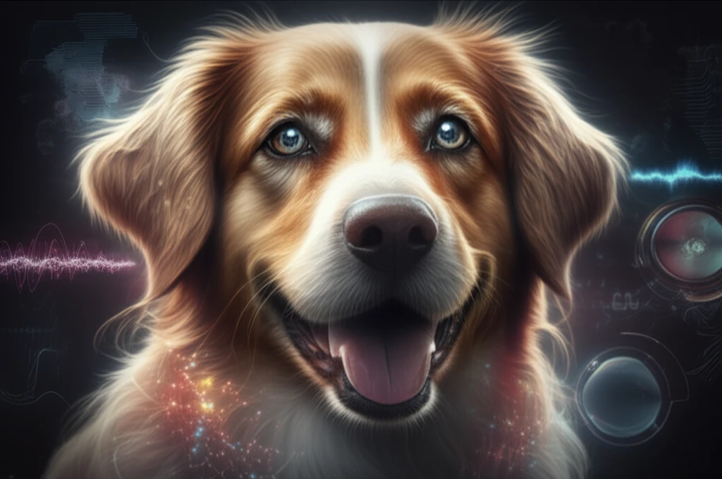
Dog with clear, sparkling eyes after successful cataract treatment.

Shedding Light on Cataracts in Dogs: An In-Depth Look at Diagnosis and Treatment

"Uncover the latest advancements in ultrasonography for diagnosing canine cataracts and how these insights are improving surgical outcomes."


Cataracts are a leading cause of blindness in dogs, often requiring surgical intervention to restore vision. Understanding the nature and extent of the cataract is crucial for successful treatment, making accurate diagnostic tools essential. One such tool, ultrasonography, has proven invaluable in assessing the lens and planning the surgical approach.

Ultrasonography offers a non-invasive way to visualize the internal structures of the eye, providing detailed images that help veterinarians determine the cataract's size, location, and density. This information is vital for surgical planning, especially when using phacoemulsification, a common technique for cataract removal. By understanding the cataract's characteristics beforehand, surgeons can tailor their approach, potentially improving outcomes and reducing complications.

This article explores the use of ultrasonography in diagnosing and managing senile cataracts in dogs. We'll delve into how this imaging technique aids in pre-surgical assessment, improves surgical precision, and ultimately enhances the quality of life for our canine companions.

Why is Early Diagnosis Key for Canine Cataract Treatment?

Dog with clear, sparkling eyes after successful cataract treatment.

Early diagnosis of cataracts in dogs is crucial for several reasons, impacting both the success of treatment and the overall well-being of the animal. Identifying cataracts early allows for timely intervention, which can prevent further vision loss and improve the chances of a successful surgical outcome.

Here's why early diagnosis matters:

  • Preventing Complete Vision Loss: Cataracts worsen over time, leading to complete blindness if left untreated. Early detection allows for intervention before significant vision loss occurs.
  • Improving Surgical Success Rates: Surgery is often more successful in the early stages of cataract development. The lens is typically softer and easier to remove, reducing the risk of complications.
  • Reducing Inflammation and Discomfort: Advanced cataracts can cause inflammation and discomfort in the eye. Early treatment can alleviate these symptoms and improve the dog's quality of life.
  • Ruling Out Other Eye Conditions: Early diagnosis allows veterinarians to rule out other underlying eye conditions that may be contributing to vision problems.
  • Better Post-Operative Recovery: Dogs undergoing cataract surgery in the early stages tend to experience quicker and smoother recovery periods.
By recognizing the importance of early diagnosis, pet owners can work with their veterinarians to monitor their dog's eye health and take proactive steps to preserve their vision.

Empowering Pet Owners Through Knowledge

Understanding the role of ultrasonography in diagnosing canine cataracts empowers pet owners to make informed decisions about their dog's eye care. By embracing proactive monitoring and early intervention, we can help our beloved companions maintain clear vision and a high quality of life.

About this Article -

This article was crafted using a human-AI hybrid and collaborative approach. AI assisted our team with initial drafting, research insights, identifying key questions, and image generation. Our human editors guided topic selection, defined the angle, structured the content, ensured factual accuracy and relevance, refined the tone, and conducted thorough editing to deliver helpful, high-quality information.See our About page for more information.

This article is based on research published under:

DOI-LINK: 10.1590/s0102-09352011000500010, Alternate LINK

Title: Ultrasonographic Analysis Of Senile Cataractous Lens Of Dogs And Its Correlation To Phacoemulsification

Subject: General Veterinary

Journal: Arquivo Brasileiro de Medicina Veterinária e Zootecnia

Publisher: FapUNIFESP (SciELO)

Authors: B.C Martins, A.P Ribeiro, J.P.D Ortiz, C.B.S Lisbão, A.L.G Souza, D Brooks, J.L Laus

Published: 2011-10-01

Everything You Need To Know

1

What are cataracts in dogs and why is it important to address them?

Cataracts in dogs occur when the lens of the eye becomes cloudy, leading to impaired vision and potential blindness. This condition often necessitates surgical intervention to restore sight. The use of diagnostic tools like ultrasonography are vital for assessing the cataract's characteristics, which helps with planning effective treatment.

2

What is ultrasonography and why is it useful in the context of cataracts?

Ultrasonography is a non-invasive imaging technique that allows veterinarians to visualize the internal structures of the eye. This is significant because it provides detailed information about the cataract's size, location, and density. This information is crucial for pre-surgical assessment and surgical planning, especially when using a technique like phacoemulsification to remove the cataract. By using ultrasonography, surgeons can tailor their surgical approach, potentially improving outcomes and reducing complications.

3

Why is it so important to diagnose cataracts in dogs early?

Early diagnosis of cataracts in dogs is important because it allows for timely intervention, which can prevent further vision loss and improve the chances of a successful surgical outcome. Early detection means intervention can occur before significant vision loss, and surgery is often more successful when the lens is softer and easier to remove, reducing the risk of complications. Early treatment can also alleviate inflammation and discomfort, and lead to quicker and smoother recovery periods.

4

What is Phacoemulsification and what is its relevance to cataract surgery?

Phacoemulsification is a common surgical technique used for cataract removal in dogs. It involves using ultrasound energy to break up the cataract into small pieces, which are then suctioned out of the eye. Ultrasonography is used in the planning phase.

5

What are senile cataracts and how is ultrasonography relevant to them?

Senile cataracts are cataracts that develop in older dogs as a result of aging. Ultrasonography plays a crucial role in diagnosing and managing senile cataracts by providing detailed images of the lens, which helps veterinarians assess the cataract's characteristics and plan the appropriate surgical approach. This is important because it enables more precise surgical intervention, potentially improving outcomes and enhancing the dog's quality of life.

Newsletter Subscribe

Subscribe to get the latest articles and insights directly in your inbox.