See Your Cells in Action: Voltage Imaging Unveiled
"Discover how genetically encoded voltage indicators (GEVIs) light up cellular communication, offering new insights into brain activity and beyond."
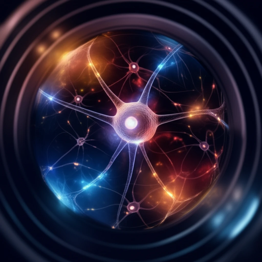
Imagine being able to watch the electrical conversations happening inside your cells. That's the promise of voltage imaging, a revolutionary technique that allows scientists to visualize changes in membrane potential—the driving force behind cellular communication. This article explores how genetically encoded voltage indicators (GEVIs) are making this possible, opening new doors in neuroscience and beyond.
Traditional methods for studying electrical activity in cells have limitations. Organic voltage-sensitive dyes, used for decades, lack specificity and can be difficult to apply to certain cell types. GEVIs offer a more targeted approach. By genetically engineering cells to express fluorescent proteins that respond to voltage changes, researchers can observe activity in specific cell populations with unprecedented precision.
This article will guide you through the principles of voltage imaging using GEVIs, highlighting the different types of sensors available and the experimental setup required. Whether you're a seasoned researcher or just curious about the inner workings of cells, this overview will provide a solid foundation for understanding this exciting field.
What are Genetically Encoded Voltage Indicators (GEVIs)?
GEVIs are essentially fluorescent reporters that change their brightness or color in response to changes in the electrical potential across a cell's membrane. They consist of a voltage-sensing domain linked to one or more fluorescent proteins. When the membrane potential changes, the voltage-sensing domain shifts, altering the fluorescent protein's properties. This allows researchers to 'see' the electrical activity of the cell.
- Single Fluorescent Protein (FP) GEVIs: These use a single fluorescent protein that changes its fluorescence intensity in response to voltage changes.
- Förster Resonance Energy Transfer (FRET)-based GEVIs: These use two fluorescent proteins, a donor and an acceptor. Voltage changes alter the distance or orientation between the proteins, affecting the efficiency of energy transfer (FRET) and changing the relative fluorescence of the donor and acceptor.
- Hybrid GEVIs: These combine a membrane-anchored fluorescent protein with a separate quenching molecule that responds to voltage.
The Future of Seeing Electricity
Voltage imaging with GEVIs is a rapidly evolving field with immense potential. As new and improved GEVIs are developed, and imaging techniques become more sophisticated, we can expect even more detailed insights into the electrical dynamics of cells and circuits. This will not only advance our understanding of fundamental biological processes but also pave the way for new diagnostic and therapeutic strategies for a wide range of neurological and other disorders.