
Securing Rectal Cancer Surgery: How a Nerve Landmark Can Prevent Urethral Injury

"A novel surgical technique uses the autonomic nerve to identify the prostate gland during transanal total mesorectal excision, minimizing the risk of urethral damage."
In rectal cancer surgery, protecting the autonomic nerve while achieving complete tumor removal is paramount. This ensures both oncological control and preservation of crucial bodily functions. Laparoscopic surgery, while beneficial, can pose challenges, especially in male patients with obesity or a narrow pelvis, due to limited visibility within the deep pelvic region. This can increase the risk of incomplete mesorectal excision and less favorable oncological outcomes.
Transanal total mesorectal excision (taTME) has emerged as a promising technique to overcome these limitations, offering improved access and visualization. However, it also carries an inherent risk of iatrogenic urethral injury in men, with reported frequencies ranging from 0% to 6.7% in single-center studies and 0.7% in the Low Rectal Cancer Development program.
Urethral injury typically occurs during anterior dissection near the preprostatic urethra, often because surgeons lack sufficient awareness of the intricate anatomy involving the neurovascular bundle of Walsh, the prostate gland, and the rectourethral muscle. Addressing this critical challenge is essential for improving patient outcomes in rectal cancer surgery.
Rare But Significant: Incidence of Male Urethral Injury
Male urethral injury during rectal cancer surgery is rare but significant, according to a comprehensive study published in Cancers (MDPI) on October 12, 2023. Urethral complications predominantly result from damage to the membranous urethra during abdominoperineal resection (the Miles procedure) or transanal total mesorectal excision (TaTME). Classical studies estimate the incidence of urethral injury during abdominoperineal resection in males at about 2%, though data on such injury in males remain scant. To address this gap, the authors used a three-pronged design—cadaveric dissection, retrospective MRI analysis, and clinical cases—to describe the anatomical relations of the male urethra, noting that scant information is available about the distances between the rectourethral space and neighboring structures.
Anatomic Mapping to Guide APR and TaTME
The reference study combined cadaveric dissection, retrospective MRI analysis, and clinical cases to achieve a better understanding of the male urethra and to prevent urethral injury during rectal cancer surgery. As its Simple Summary states, urethral injuries in men undergoing abdominoperineal resection or TaTME for rectal cancer are uncommon but devastating, and the review of images obtained through these methods was intended to clarify those anatomical relations. The study notes that scant information is available about the distances between the rectourethral space and neighboring structures, which limits current surgical guidance. By cross-referencing multiple lines of evidence, the authors aim to give surgeons a more precise anatomical basis for the anterior dissection performed in these procedures.
Foundations in Pelvic Surgical Anatomy
Preventing urethral injury in rectal cancer surgery has long rested on anatomical knowledge of the male pelvis, although the specific historical milestones are not detailed in the material reviewed here. The abdominoperineal resection, known as the Miles procedure, and the later arrival of transanal approaches each focused surgical attention on the membranous urethra lying in the anterior field. Cadaveric study has historically been a foundational method for mapping this region and the rectourethral space. Without a documented timeline in the available sources, this history is best characterized as a gradual refinement of anatomical understanding rather than a series of discrete breakthroughs.
Autonomic Nerve as a Guide: A Step-by-Step Surgical Technique
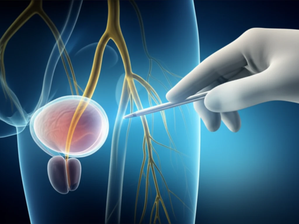
To address the risk of urethral injury, a novel surgical technique has been developed that uses the autonomic nerve as a landmark to identify the prostate gland and ensure safe dissection of the male rectourethral muscle. This approach aims to enhance precision and minimize the potential for damage during taTME.
- Initial Dissection: Circumferential rectal dissection begins at the anorectal ring, proceeding posteriorly and cranially within the avascular presacral plane.
- Nerve Identification: Dissection from the posterior wall towards the left side facilitates recognition of the rise of the fourth pelvic splanchnic nerve (S4). This nerve serves as a critical landmark.
- Prostate Identification: By tracing the autonomic nerves anteriorly while carefully identifying S4 and the neurovascular bundle, the prostate gland can be accurately located before dissecting the rectourethral muscle.
- Safe Muscle Dissection: Using this nerve-guided approach, the rectourethral muscle can be safely cut while clearly visualizing the prostate gland, thus avoiding urethral injury. This also allows for precise selection of the cutoff line of the rectourethral muscle based on individual patient anatomy.
Emerging Evidence and Continuing Review
Recent work in this area centers on better defining the anatomy of the rectourethral region in order to reduce the uncommon but serious risk of urethral injury during rectal cancer surgery. Published reviews continue to emphasize the potential for significant short-term complications and long-term effects when the urethra is damaged. A research design combining dissection, imaging, and clinical series is emerging as a favored approach for producing usable anatomical guidance. Findings at this stage should still be treated as incremental, since much of the available evidence is descriptive rather than experimental.
Challenges and Unanswered Questions
One persistent challenge is the relative scarcity of data on male urethral injury, which limits how confidently incidence and risk can be stated. The complexity of the anterior surgical plane means that even careful dissection carries a residual risk of injuring the membranous urethra. None of the material reviewed here identifies a single best strategy, and approaches vary with the type of procedure and the surgeon's experience. Claims about eliminating this complication should be viewed cautiously, because the underlying evidence is limited and largely descriptive.
Comparing Surgical Approaches
Comparative discussion in this area generally contrasts open techniques such as abdominoperineal resection with more recent transanal approaches like TaTME, both of which place the membranous urethra at risk. Each approach offers a different view of the rectourethral space, and no single method in the material reviewed is presented as uniformly superior. Cross-referencing dissection findings with imaging appears valuable because each information source covers different gaps in understanding. Without head-to-head outcome data, such comparisons remain anatomical and methodological rather than definitive clinical evidence.
Reducing Risks, Improving Outcomes
By adopting this surgical technique, surgeons can significantly reduce the risk of urethral injury during taTME, a critical step towards improving patient safety and outcomes in rectal cancer surgery.
An Emerging Consensus on Anatomical Awareness
Where commentary is available, it converges on a central theme: urethral injury during rectal cancer surgery is rare but should not be treated as negligible, given the potential for significant short-term complications and long-term effects. Understanding the precise anatomical relations of the male urethra—particularly the distances within the rectourethral space—is presented as the key to prevention. There is growing support for combining multiple lines of evidence, such as dissection and imaging, to give surgeons dependable landmarks. This is best read as a developing consensus rather than a settled clinical protocol.
Toward Safer Anterior Dissection
Looking ahead, the clearest opportunity appears to be generating more systematic anatomical data that can translate directly into surgical guidance. If larger clinical series can document the distances between the rectourethral space and neighboring structures, they would likely inform training and operative checkpoints. Whether new imaging tools or standardized dissection techniques will accelerate progress remains unclear from the material reviewed here. Any projected decline in injury rates is speculative at this stage.
Systemic Gaps in Study and Training
A broader obstacle is that rare complications are inherently difficult to study, since individual centers accumulate too few cases to draw firm conclusions. The emphasis on anatomical teaching in surgical training reflects a systemic response, though its effectiveness is not quantified in the available material. Limited data on male urethral injury also constrain how well risk can be communicated to surgeons in training. These systemic factors, while plausible, are presented here as observations rather than as findings established by the reviewed sources.
A Case of Immediate Reconstruction and Four Years of Follow-Up
A case report describes a 57-year-old man with advanced rectoanal cancer who underwent laparoscopy-assisted abdominoperineal resection, during which the anterior rectal wall dissection resulted in a membranous urethral injury. The authors note that urethral injury, although occurring at a measurable frequency during rectal surgery, remains underrecognized by urologists. Immediate urethral reconstruction performed during the same cancer operation preserved urinary function, with no evidence of stricture or incontinence through four years of follow-up. The successful long-term outcome supports immediate reconstruction as a viable response when injury is recognized intraoperatively.
The key lies in using the autonomic nerve as a reliable landmark to guide dissection and accurately identify the prostate gland. This approach enhances precision and control, minimizing the potential for iatrogenic damage.
Further research and widespread adoption of this technique could lead to a new standard of care in rectal cancer surgery, ensuring both effective tumor removal and preservation of vital anatomical structures.