Say Goodbye to Blurry Endoscopies: The AI Tech Sharpening the Future of Surgery
"Discover how a novel image enhancement technique is poised to revolutionize minimally invasive surgery, offering clearer visuals and safer procedures."
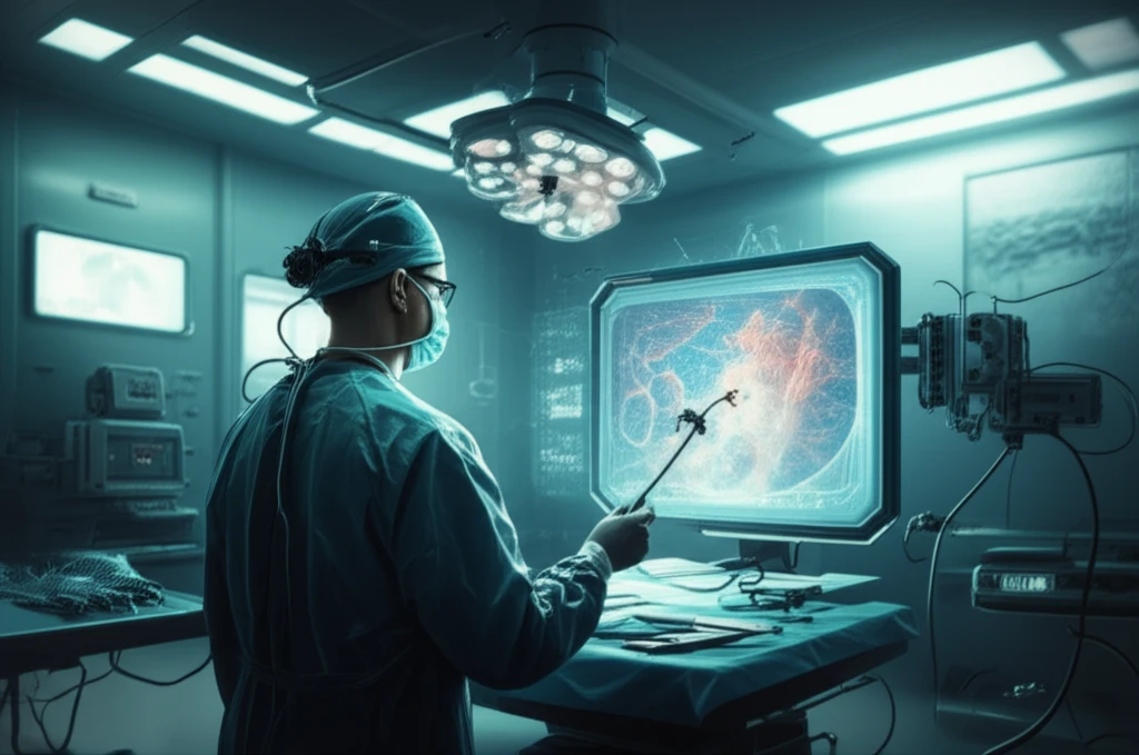
Minimally invasive surgery has been revolutionized through stereoscopic endoscopes, which provide surgeons with the ability to visualize organ surfaces and use surgical tools with increasing accuracy. A persistent obstacle in endoscopic surgery is the issue of insufficient and irregular light sources. This issue significantly affects image quality and occasionally causes surgical tools to be hardly apparent in low-light environments.
Images taken in low-light conditions are more likely to have a low signal-to-noise ratio and metrication artifacts brought on by quantization errors. Consequently, conventional image enhancement techniques frequently worsen noise in low-light areas, which compromises image clarity and surgical precision. Current methods frequently amplify existing noise during this process, reducing their efficacy.
This article explores a groundbreaking method designed to improve endoscopic image quality by identifying distinct illumination zones and tailoring enhancement strategies according to desired visual results. This novel technique dramatically reduces noise amplification during image processing, especially in previously challenging low-light circumstances, in contrast to existing enhancement approaches. This could be especially useful in preventing damage to sensitive organs like the liver and spleen.
How Does AI Enhance Endoscopic Images?
The key lies in a refined approach to image processing, which addresses typical challenges such artifacts, noise, and uneven illumination. The method begins by evaluating the source and nature of the image's lighting environment, and then tailors processes to enhance image clarity, reduce artifacts, and limit noise. This process relies on several steps:
- Illumination Region Identification: The algorithm begins by segmenting the endoscopic image into three distinct regions based on lighting conditions: well-lit, low-light, and lossy (extremely low light with significant detail loss). This is achieved by analyzing the V-space element from the HSV (Hue, Saturation, Value) transformation of the image, using predefined thresholds to categorize each pixel.
- Base and Detail Layer Decomposition: After identifying the illumination regions, the image is divided into a base layer, representing smooth luminance variations, and a detail layer, capturing local contrasts and fine details. This decomposition is performed using an edge-preserving smoothing filter, such as the tree filter, which efficiently separates the image components while preserving important structural details.
- Enhancement Factor Application: Enhancement factors are then applied to both the base and detail layers, tailored to each illumination region. The well-lit regions are largely preserved to maintain their natural appearance, while the low-light regions undergo a gamma correction-like enhancement to improve luminance. In the lossy regions, a linear gain is applied to suppress noise amplification, ensuring that only genuine details are enhanced.
- Adaptive Smoothing: To further refine the enhancement process, the pseudo enhancement factors for both base and detail layers are smoothed using Gaussian kernels. This step helps to enforce a piece-wise linear assumption of the illumination, reducing artifacts and ensuring a seamless transition between different enhancement levels.
- Image Reconstruction: Finally, the enhanced base and detail layers are combined to produce the final enhanced image. This reconstructed image exhibits improved visibility in low-light areas, reduced noise and artifacts, and an overall natural appearance.
The Future of Surgical Imaging
The innovative method not only improves image quality but also lays the groundwork for more sophisticated image processing applications in surgery. The results show that this method improves endoscopic images by preserving contrast and color in well-lit areas while improving low-light visibility. This leads to more natural-looking images with less noise. This method outperforms existing technologies, increasing its effectiveness in endoscopic procedures.