Futuristic operating room with holographic liver projection.

Revolutionizing Liver Surgery: The Rise of Real-Time Navigation

"Discover how a novel automatic registration system is making liver resections safer, faster, and more accurate, benefiting both surgeons and patients."


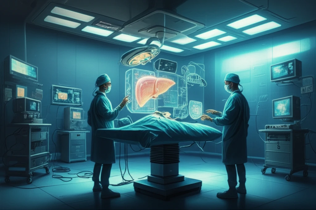
Liver resection, a standard treatment for primary and secondary liver malignancies, has seen remarkable advancements. Three-dimensional (3D) simulation software now enables more precise preoperative planning, often referred to as virtual hepatectomy. However, the challenge remains in seamlessly transferring this pre-operative plan to the actual intraoperative setting.

Surgeons must not only understand the preoperative plan but also intuitively identify the optimal liver transection line and locate critical vessels buried within the liver. To address this, intraoperative ultrasonography (IOUS) has been utilized since the 1980s, yet it requires extensive knowledge of liver anatomy and considerable experience to interpret the images effectively.

Real-time virtual sonography (RVS) has emerged as a valuable tool, aiding in the interpretation of ultrasound images by providing synchronized two-dimensional (2D) computed tomography (CT) images alongside ultrasound views. This technology allows surgeons to visualize target vessels and planned transection lines marked with colors using 3D simulation software. A recent breakthrough involves a novel automatic registration system that simplifies and accelerates the alignment of CT and IOUS images, potentially enhancing the precision and accessibility of liver surgery.

How Does the New RVS System Work?

Futuristic operating room with holographic liver projection.

The enhanced RVS navigation system integrates intraoperative ultrasonography with electromagnetic tracking technology. It features an ultrasound scanner with a specialized intraoperative ultrasound probe, an electromagnetic tracking device, and a workstation for real-time analysis. During surgery, an electromagnetic transmitter is positioned near the patient, and a sterilized sensor is attached to the ultrasound probe, enabling precise tracking of the probe's location and orientation.

Before the surgery, contrast-enhanced CT images with a 1-mm slice thickness are obtained, and the liver's anatomical structures, including the portal and hepatic veins, are extracted using 3D simulation software. An experienced liver surgeon identifies key hepatic venous tributaries, such as V8 and V5, which drain segments S8 and S5, respectively. All reconstructed 3D and 2D CT images are then transferred to the RVS workstation.

  • Initial Point Definition: The RVS workstation begins by defining an 'initial point,' which roughly aligns the spatial position and direction of the IOUS probe. This point, located on the left liver surface, is identified in both the 2D CT images and the IOUS view.
  • Diagonal Scanning: Surgeons perform a 'diagonal scan' by moving the IOUS probe across the liver surface, capturing a volume of ultrasound data around the bifurcation of the left and right portal veins. This scan typically covers about 15 cm of the liver surface and takes only a few seconds.
  • Automatic Registration: The acquired ultrasound volume images are reconstructed into 3D images and compared with the preoperatively obtained 3D CT images at the workstation. The system automatically matches these two sets of 3D images, adjusting the position and orientation of the CT images to achieve accurate multiplanar reconstruction.
This automated process significantly reduces the reliance on manual registration, which requires considerable experience and expertise in interpreting IOUS images and matching anatomical points between 2D CT images and IOUS views. The new system streamlines the workflow and improves accuracy, making it more accessible to surgeons with varying levels of experience.

The Future of Liver Surgery

The RVS system with automatic registration represents a significant step forward in liver surgery. By providing quick, easy, and accurate image registration, this technology enhances the precision and efficiency of liver resections. While challenges remain, such as compensating for liver deformation and minimizing positional errors, the RVS system has the potential to improve surgical outcomes and expand the use of IOUS in liver surgery, ultimately benefiting patients and surgeons alike.

About this Article -

This article was crafted using a human-AI hybrid and collaborative approach. AI assisted our team with initial drafting, research insights, identifying key questions, and image generation. Our human editors guided topic selection, defined the angle, structured the content, ensured factual accuracy and relevance, refined the tone, and conducted thorough editing to deliver helpful, high-quality information.See our About page for more information.

This article is based on research published under:

DOI-LINK: 10.1007/s00268-017-4210-5, Alternate LINK

Title: Feasibility Of Intraoperative Navigation For Liver Resection Using Real-Time Virtual Sonography With Novel Automatic Registration System

Subject: Surgery

Journal: World Journal of Surgery

Publisher: Springer Science and Business Media LLC

Authors: Takeshi Takamoto, Yoshihiro Mise, Shouichi Satou, Yuta Kobayashi, Koui Miura, Akio Saiura, Kiyoshi Hasegawa, Norihiro Kokudo, Masatoshi Makuuchi

Published: 2017-09-06

Everything You Need To Know

1

How does the new Real-time virtual sonography (RVS) system actually function?

The new Real-time virtual sonography (RVS) system works by integrating intraoperative ultrasonography (IOUS) with electromagnetic tracking technology. It uses a specialized intraoperative ultrasound probe and an electromagnetic tracking device to precisely track the probe's location and orientation. Before surgery, 3D simulation software is used to extract the liver's anatomical structures from contrast-enhanced CT images. During surgery, the RVS workstation defines an 'initial point' on the liver and surgeons perform a 'diagonal scan' to capture ultrasound data. The system then automatically matches the ultrasound data with the preoperative 3D CT images, improving the accuracy of the surgery.

2

Why is the new RVS system important for liver surgery?

The importance of the RVS system is that it streamlines the workflow and improves the accuracy of liver resections. By automating the alignment of CT and IOUS images, the new RVS system reduces the reliance on manual registration, which requires significant experience in interpreting IOUS images and matching anatomical points. This makes the technology more accessible to surgeons with varying levels of experience. It enhances the precision and efficiency of liver resections, which is a standard treatment for liver malignancies.

3

What is the role of Intraoperative ultrasonography (IOUS) in the context of the new technology?

Intraoperative ultrasonography (IOUS) is a crucial part of the RVS system because it provides real-time images of the liver during surgery, allowing surgeons to visualize critical structures such as vessels and planned transection lines. However, IOUS images can be difficult to interpret. The RVS system uses 2D computed tomography (CT) images alongside ultrasound views to assist in the interpretation of the ultrasound images, which is a significant improvement over the traditional methods.

4

How is 3D simulation software used with this new technology?

3D simulation software is used before the surgery to create a detailed preoperative plan, including a 'virtual hepatectomy'. This helps surgeons identify the optimal liver transection line and locate critical vessels. The RVS system uses the 3D CT images created by the software to match with the IOUS images to provide real-time guidance. The preoperative planning ensures that surgeons have a comprehensive understanding of the liver's anatomy before the actual procedure, ultimately improving the precision of the surgery.

5

What are the potential implications of this new RVS system for the future of liver surgery?

The implications of the new RVS system are far-reaching. It has the potential to improve surgical outcomes by making liver resections safer, faster, and more accurate. This could expand the use of IOUS in liver surgery. While challenges like liver deformation and positional errors remain, the advancements in technology have made complex procedures more manageable. It ultimately benefits both surgeons by streamlining their workflow and patients by increasing the chances of successful liver surgery.

Newsletter Subscribe

Subscribe to get the latest articles and insights directly in your inbox.