Illustration of a fibrolipoma within a human chest cavity

Rare Chest Mass? A Fibrolipoma Case Study & What It Means For You

"Uncommon tumors, cutting-edge surgery, and the importance of early detection. A deep dive into a rare intrathoracic fibrolipoma case."


In the realm of medical anomalies, lipomas—benign tumors composed of fat cells—are relatively common. However, these fatty growths sometimes present in unexpected forms. One such variant is the fibrolipoma, a type of lipoma containing a significant proportion of fibrous tissue and blood vessels. While lipomas are already diverse, intrathoracic fibrolipomas, located within the chest cavity, are remarkably rare. To put things in perspective, they are so rare that, to the best of medical knowledge, only three cases have been documented.

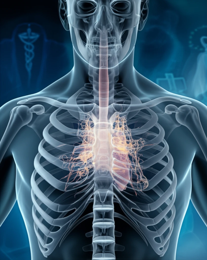
Recently, a team of surgeons successfully resected an intrathoracic fibrolipoma using complete thoracoscopic surgery. This achievement marks a significant milestone in treating such rare conditions. The patient, a 51-year-old female, presented with a left intrathoracic mass discovered during a routine medical checkup. What followed was a carefully orchestrated medical intervention that underscores the importance of proactive health monitoring and cutting-edge surgical techniques.

This article delves into the details of this extraordinary case, exploring the nature of fibrolipomas, the diagnostic process, the surgical approach, and the implications for future medical practice. It also highlights why such cases, though rare, are crucial in advancing medical knowledge and patient care.

What is a Fibrolipoma?

Illustration of a fibrolipoma within a human chest cavity

Lipomas, generally composed of adipocytes (fat cells), can sometimes include mesodermal components such as fibrous tissues and blood vessels. Fibrolipomas are a variant where fibrous tissue is a major component. These tumors are benign, but their location can sometimes cause complications.

While lipomas account for about 20% of all benign soft tissue tumors, fibrolipomas make up a tiny fraction—approximately 0.03% [4]. They can occur in various body parts, including subcutaneous and muscular tissues. However, intrathoracic fibrolipomas, found within the chest cavity, are exceptionally rare. This rarity makes each case significant for medical research and clinical understanding.

  • Rarity: Intrathoracic fibrolipomas are exceptionally rare, with only a handful of cases reported.
  • Composition: These tumors consist of both fat and fibrous tissue, making them distinct from regular lipomas.
  • Location: Occurring in the chest cavity, they can potentially affect surrounding organs and tissues.
The recent case report underscores the critical role of advanced imaging techniques such as Computed Tomography (CT) and Positron Emission Tomography (PET) in identifying and characterizing such lesions. These tools aid in differentiating benign tumors from malignant ones, guiding appropriate treatment strategies. Understanding these factors is vital for healthcare professionals in diagnosing and managing similar cases.

Why This Case Matters

This successful resection of an intrathoracic fibrolipoma using complete thoracoscopic surgery marks an important advancement. The tumor was asymptomatic and relatively small, discovered during a routine checkup. This early detection enabled a less invasive surgical approach, highlighting the importance of regular medical screenings. Though fibrolipomas are benign, careful observation and follow-up are essential due to the possibility of recurrence [7]. Continuous monitoring and proactive healthcare management remain crucial for ensuring long-term well-being.

About this Article -

This article was crafted using a human-AI hybrid and collaborative approach. AI assisted our team with initial drafting, research insights, identifying key questions, and image generation. Our human editors guided topic selection, defined the angle, structured the content, ensured factual accuracy and relevance, refined the tone, and conducted thorough editing to deliver helpful, high-quality information.See our About page for more information.

This article is based on research published under:

DOI-LINK: 10.1186/s13019-018-0808-4, Alternate LINK

Title: Intrathoracic Fibrolipoma Resected Using Complete Thoracoscopic Surgery: A Case Report

Subject: Cardiology and Cardiovascular Medicine

Journal: Journal of Cardiothoracic Surgery

Publisher: Springer Science and Business Media LLC

Authors: Satoshi Kamata, Itaru Ishida, Yuyo Suzuki, Takehiro Yamada, Hiroshi Yaegashi, Hiroyuki Oura

Published: 2018-11-14

Everything You Need To Know

1

What exactly is a fibrolipoma, and how does it differ from a regular lipoma?

A fibrolipoma is a type of benign tumor that, like a regular lipoma, contains fat cells. However, unlike regular lipomas, a fibrolipoma contains a significant proportion of fibrous tissue and blood vessels in addition to fat cells (adipocytes). While lipomas are composed primarily of fat, fibrolipomas have a substantial amount of fibrous tissue, making them distinct. Their location, particularly when intrathoracic, can sometimes cause complications due to the surrounding organs and tissues in the chest cavity. Understanding the composition and potential location is crucial for diagnosis and management.

2

Why is the intrathoracic fibrolipoma case considered so rare and significant in the medical field?

The intrathoracic fibrolipoma case is considered exceptionally rare because these tumors, located within the chest cavity, are infrequently documented. To the best of medical knowledge, only a few cases have been reported. Each instance provides valuable insights into the nature, diagnosis, and treatment of such unusual conditions. The successful resection using complete thoracoscopic surgery in the recent case marks a significant advancement in treating these rare conditions and underscores the importance of proactive health monitoring and cutting-edge surgical techniques in managing rare medical anomalies.

3

How are advanced imaging techniques like CT and PET scans crucial in diagnosing fibrolipomas, and what role do they play in treatment planning?

Advanced imaging techniques such as Computed Tomography (CT) and Positron Emission Tomography (PET) scans are critical in identifying and characterizing fibrolipomas. These tools help differentiate benign tumors like fibrolipomas from potentially malignant ones, guiding appropriate treatment strategies. CT scans provide detailed anatomical images to determine the size and location of the tumor, while PET scans can assess the metabolic activity to rule out malignancy. This differentiation is essential for planning the appropriate surgical approach, such as the complete thoracoscopic surgery used in the recent case. Without these imaging techniques, accurate diagnosis and treatment planning would be significantly more challenging.

4

What are the implications of early detection and proactive health monitoring in managing conditions like intrathoracic fibrolipomas?

Early detection and proactive health monitoring are vital in managing conditions like intrathoracic fibrolipomas. In the recent case, the tumor was discovered during a routine medical checkup while asymptomatic. This early detection enabled a less invasive surgical approach, specifically complete thoracoscopic surgery, leading to a successful outcome. Regular medical screenings and proactive healthcare management are crucial because, although fibrolipomas are benign, careful observation and follow-up are essential due to the possibility of recurrence. Continuous monitoring ensures long-term well-being and allows for timely intervention if any changes or complications arise.

5

Given the rarity of intrathoracic fibrolipomas, what future research or clinical practices could improve our understanding and treatment of these tumors?

Given the rarity of intrathoracic fibrolipomas, several avenues of research and clinical practice could enhance our understanding and treatment of these tumors. Establishing a national or international registry of fibrolipoma cases could facilitate collaborative research and data sharing among medical professionals. Further investigation into the genetic and molecular characteristics of fibrolipomas may reveal potential therapeutic targets. Additionally, refining minimally invasive surgical techniques, such as complete thoracoscopic surgery, and developing standardized follow-up protocols can improve patient outcomes. Increased awareness and education among healthcare providers about the possibility of fibrolipomas in unusual locations can also lead to earlier diagnosis and intervention.

Newsletter Subscribe

Subscribe to get the latest articles and insights directly in your inbox.