Surreal illustration of lung and plasma cells symbolizing diagnostic challenges in cancer.

Rare Case Study: When Lung Cancer Mimics Plasma Cell Myeloma

"Unveiling the Diagnostic Challenges and Innovative Solutions in a Unique Cancer Presentation"


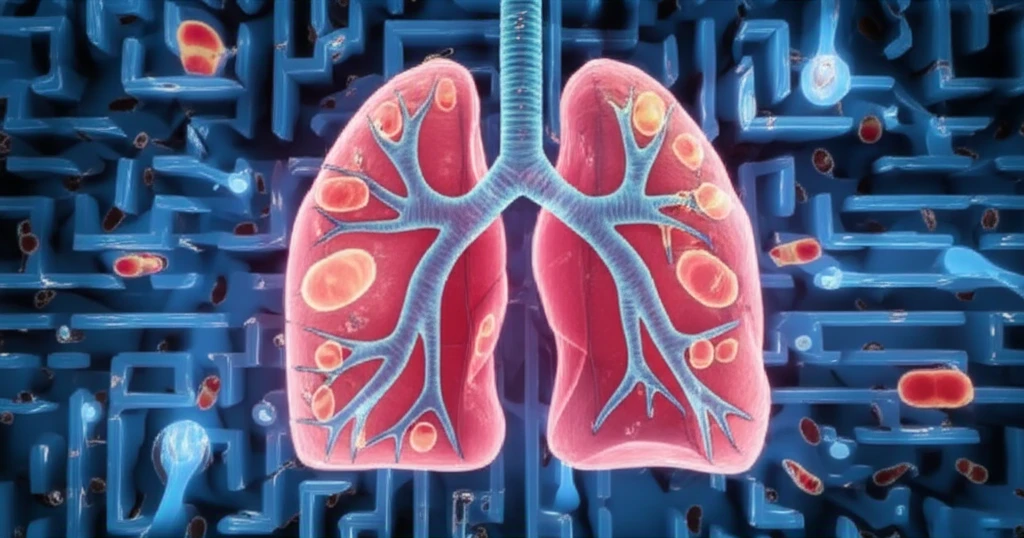
Plasma cell myeloma (PCM) is a cancer of the plasma cells, which are a type of white blood cell responsible for producing antibodies. Typically, PCM is found in the bone marrow, where these cells reside and multiply. However, in rare instances, PCM can manifest outside the bone marrow in what is known as extramedullary plasmacytoma. When this occurs in the lungs, it can create a diagnostic puzzle, mimicking other lung conditions, particularly lung cancer.

Diagnosing PCM in the lungs is especially challenging because the symptoms and initial findings can closely resemble those of lung cancer. Both conditions may present with lung masses, pleural effusions (fluid around the lungs), and even bone involvement. This overlap necessitates a thorough and nuanced diagnostic approach to ensure patients receive the correct and timely treatment.

This article will delve into a fascinating case where PCM initially masqueraded as lung cancer, causing a diagnostic dilemma. We’ll explore the steps taken to unravel the mystery, the innovative diagnostic tools used, and the lessons learned from this unusual presentation. Understanding such rare cases is crucial for clinicians to enhance their diagnostic acumen and improve patient outcomes.

The Case Presentation: A Diagnostic Puzzle

Surreal illustration of lung and plasma cells symbolizing diagnostic challenges in cancer.

A 59-year-old Korean woman presented to the hospital with complaints of anorexia and weight loss that had persisted for six weeks. A routine check-up at a local clinic had incidentally revealed a lung mass, prompting further investigation. The patient had no history of smoking or asbestos exposure, common risk factors for lung cancer.

Initial blood tests revealed some abnormalities, including mild anemia (Hb 8.9 g/dL) and slightly reduced white blood cell count (3.8×10⁹/L). Biochemical tests showed normal kidney function but elevated lactate dehydrogenase (LDH), an enzyme that can indicate tissue damage or cancer.

  • Chest computed tomography (CT) scans showed a lung mass in the left lower lobe with a lobulated contour.
  • Bony destructive soft tissue masses were present in the left ribs.
  • Multifocal pre/paravertebral mass lesions were noted, especially at the T9-L1 level.
  • Pleural effusions (fluid accumulation around the lungs) were observed.
Based on these initial radiologic findings, the primary suspicion was lung cancer with pleural metastasis. However, the possibility of PCM was also considered in the differential diagnosis.

Key Takeaways and the Importance of Vigilance

This case underscores the critical importance of maintaining a broad differential diagnosis and utilizing advanced diagnostic techniques when faced with atypical presentations of common diseases. While lung cancer remains a primary concern in patients presenting with lung masses and pleural effusions, clinicians should be vigilant about considering rarer conditions like PCM, especially when initial findings are not entirely consistent with typical lung cancer presentations. By employing a combination of imaging, cytological examination, electrophoresis, and free light chain assays, healthcare professionals can improve their ability to accurately diagnose and effectively manage complex cases of PCM and other challenging malignancies. This ultimately leads to better outcomes and improved quality of life for patients.

About this Article -

This article was crafted using a human-AI hybrid and collaborative approach. AI assisted our team with initial drafting, research insights, identifying key questions, and image generation. Our human editors guided topic selection, defined the angle, structured the content, ensured factual accuracy and relevance, refined the tone, and conducted thorough editing to deliver helpful, high-quality information.See our About page for more information.

This article is based on research published under:

DOI-LINK: 10.3343/alm.2013.33.3.225, Alternate LINK

Title: Plasma Cell Myeloma Initially Presenting As Lung Cancer

Subject: Biochemistry (medical)

Journal: Annals of Laboratory Medicine

Publisher: Annals of Laboratory Medicine

Authors: Sun Young Cho, Jae-Heon Jeong, Woo-In Lee, Juhie Lee, Il Ki Hong, Jin-Tae Suh, Hee Joo Lee, Hwi-Joong Yoon, Tae Sung Park

Published: 2013-05-01

Everything You Need To Know

1

What is Plasma Cell Myeloma (PCM), and how does it typically manifest?

Plasma Cell Myeloma (PCM) is a cancer of the plasma cells, a type of white blood cell responsible for producing antibodies. Typically, PCM is found in the bone marrow, where these cells reside and multiply. However, in rare cases, PCM can manifest outside the bone marrow as an extramedullary plasmacytoma, sometimes mimicking other conditions. This case illustrates the diagnostic challenges when PCM presents in an atypical location such as the lungs. Advanced testing like electrophoresis and free light chain assays are required to differentiate PCM from other malignancies.

2

Why is diagnosing Plasma Cell Myeloma (PCM) in the lungs so difficult?

Diagnosing Plasma Cell Myeloma (PCM) in the lungs is challenging because its symptoms and initial findings often resemble those of lung cancer. Both conditions can present with lung masses, pleural effusions (fluid around the lungs), and even bone involvement. This overlap necessitates a thorough and nuanced diagnostic approach. Without advanced diagnostic techniques like electrophoresis and free light chain assays, clinicians may misdiagnose PCM as lung cancer, leading to inappropriate treatment.

3

What specific diagnostic tools are most helpful in differentiating between Plasma Cell Myeloma (PCM) and lung cancer when both present with similar symptoms?

In differentiating between Plasma Cell Myeloma (PCM) and lung cancer, specific diagnostic tools play a crucial role. Imaging techniques such as chest computed tomography (CT) scans help visualize lung masses and pleural effusions. However, cytological examinations, electrophoresis, and free light chain assays are essential for definitive diagnosis. Electrophoresis can identify abnormal protein levels indicative of PCM, while free light chain assays can detect the presence of monoclonal light chains, a hallmark of PCM. These advanced tests provide critical information that distinguishes PCM from lung cancer.

4

What were the initial findings in the presented case that led doctors to suspect lung cancer, and what eventually pointed towards Plasma Cell Myeloma (PCM)?

In the presented case, the initial findings that led doctors to suspect lung cancer included a lung mass in the left lower lobe observed on chest computed tomography (CT) scans, bony destructive soft tissue masses in the left ribs, multifocal pre/paravertebral mass lesions, and pleural effusions. These findings, combined with the patient's anorexia and weight loss, initially suggested lung cancer with pleural metastasis. However, the possibility of Plasma Cell Myeloma (PCM) was also considered in the differential diagnosis, prompting further investigation using advanced techniques like electrophoresis and free light chain assays, which ultimately confirmed the PCM diagnosis.

5

What are the implications of misdiagnosing Plasma Cell Myeloma (PCM) as lung cancer, and how can clinicians avoid this error?

Misdiagnosing Plasma Cell Myeloma (PCM) as lung cancer can lead to inappropriate and ineffective treatment, potentially worsening the patient's condition and delaying proper care. Lung cancer treatments, such as surgery or radiation, would not address the underlying plasma cell malignancy. To avoid this error, clinicians should maintain a broad differential diagnosis, especially when initial findings are not entirely consistent with typical lung cancer presentations. Employing a combination of imaging, cytological examination, electrophoresis, and free light chain assays is crucial for accurate diagnosis. Vigilance and the use of advanced diagnostic techniques are essential to differentiate PCM from other malignancies and ensure patients receive the correct treatment.

Newsletter Subscribe

Subscribe to get the latest articles and insights directly in your inbox.