Radial Scars After Breast Biopsy: When to Worry?
"Navigating the Controversy: Understanding Histology, Imaging, and Management of Radial Scars Found During Breast Core Biopsy."
Radial scars are a common, benign finding in breast tissue. Diagnosed with increasing frequency thanks to advancements in imaging technology, particularly digital tomosynthesis, these lesions often present a management dilemma.
Historically, radial scars were identified incidentally during surgery for clinically suspicious masses or breast cancer. Now, they are frequently detected through routine mammography, ultrasound, or MRI, becoming targets for image-guided biopsies.
The challenge arises after a core biopsy reveals a radial scar. Uncertainty surrounds the scar's potential to become malignant and its possible coexistence with other high-risk lesions or even breast cancer. While a single, definitive guideline is impossible, this article offers a balanced perspective and outlines reasonable management strategies.
Understanding the Pathology of Radial Scars
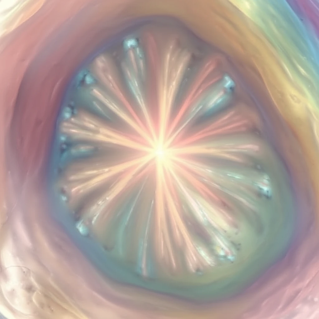
Radial scars are characterized by a central fibroelastotic core with radiating spokes of ducts and lobules. Imagine a tiny bicycle wheel where the hub is a dense, fibrous center, and the spokes are normal breast structures stretched outwards.
- Size Matters (Sort Of): Some experts differentiate radial scars (less than 1 cm) from complex sclerosing lesions (larger than 1 cm), but both share similar characteristics.
- Entrapment Illusion: Glandular elements can sometimes get trapped in the central core, mimicking tubular carcinoma under a microscope. Special staining techniques can help pathologists differentiate between benign and malignant cells.
- Common, but Not Predictive: Autopsy studies reveal radial scars in 14% to 28% of unselected female breasts. Having a radial scar doesn't necessarily increase breast cancer risk beyond that associated with any accompanying proliferative changes.
Making Informed Decisions: The Path Forward
Managing radial scars after core biopsy requires a balanced approach. Current research suggests these lesions aren't inherently premalignant, but a small percentage are upgraded to malignancy after surgical removal, likely due to sampling errors during the core biopsy.
To minimize this risk, consider these general guidelines:
<ul> <li><b>Small Scars (≤ 1.0 cm):</b> If the scar is small, well-sampled, and shows agreement between imaging and pathology, careful monitoring may suffice.</li> <li><b>Larger Scars (> 1.0 cm):</b> Excision or repeat sampling with a larger-gauge vacuum-assisted device might be necessary to minimize sampling errors.</li> <li><b>Incidental Scars:</b> Small, incidental scars found during biopsy of another lesion may only require imaging and clinical follow-up if the primary target is adequately sampled and concordant.</li> </ul> These recommendations are general and can be adapted based on individual factors such as personal history, sampling technique, and the relationship between the biopsy clip and lesion. When in doubt, surgical excision is the safest approach.