Pulmonary Lipiodol Accumulation: What to Know About This Rare TACE Complication
"Understanding the causes, symptoms, and management of pulmonary lipiodol accumulation after transarterial chemoembolization."
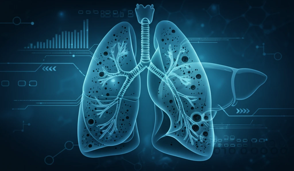
Transarterial chemoembolization (TACE) is a crucial procedure in treating hepatocellular carcinoma (HCC), a common type of liver cancer. By delivering chemotherapy directly to the tumor site, TACE effectively blocks the tumor's blood supply, slowing its growth. This targeted approach minimizes damage to healthy tissues, improving patient outcomes. However, like any medical intervention, TACE isn't without potential complications. One such complication, though rare, is pulmonary lipiodol accumulation (PLA).
PLA occurs when lipiodol, an oily contrast agent used in TACE, unintentionally accumulates in the lungs. While TACE is designed to target tumors, the accidental perfusion of lipiodol into arteriovenous shunts or the inferior phrenic artery can lead to PLA. This accumulation can manifest in various ways, sometimes accompanied by intrathoracic abnormalities. Understanding the causes, symptoms, and management of PLA is essential for both healthcare professionals and patients undergoing TACE.
This article delves into the specifics of PLA, exploring its incidence, diagnosis, and radiologic outcomes following TACE. Drawing on clinical research, we aim to provide a comprehensive overview of this rare complication, offering insights into its management and potential impact on patient recovery.
What are the Symptoms and CT Findings of Pulmonary Lipiodol Accumulation?
The symptoms and CT findings of pulmonary lipiodol accumulation (PLA) can vary, but it's essential to recognize the potential signs. While some patients may experience no noticeable symptoms, others might develop respiratory issues such as cough, fever, dyspnea (shortness of breath), or hemoptysis (coughing up blood). The severity of these symptoms can range from mild discomfort to more significant respiratory distress.
- Asymptomatic Presentation: Some individuals may show no immediate symptoms, with the accumulation only detected through imaging.
- Respiratory Symptoms: Potential symptoms include cough, shortness of breath, fever, and hemoptysis.
- CT Scan Findings: Accumulations appear as dense areas, often in the lower lobes, potentially accompanied by pleural effusion or consolidation.
Prognosis and Treatment of Pulmonary Lipiodol Accumulation
The recovery time for lipiodol accumulation is approximately 66 days. The clinical and radiological outcome of PLA without respiratory failure is promising, and conservative treatment is sufficient when lipiodol accumulation is observed in CT images after TACE.