Illustration of lungs affected by tapeworm cysts

Pulmonary Hydatid Cysts: What You Need to Know About This Rare Lung Infection

"Understanding the causes, symptoms, and treatment of pulmonary hydatid cysts, a parasitic infection affecting the lungs."


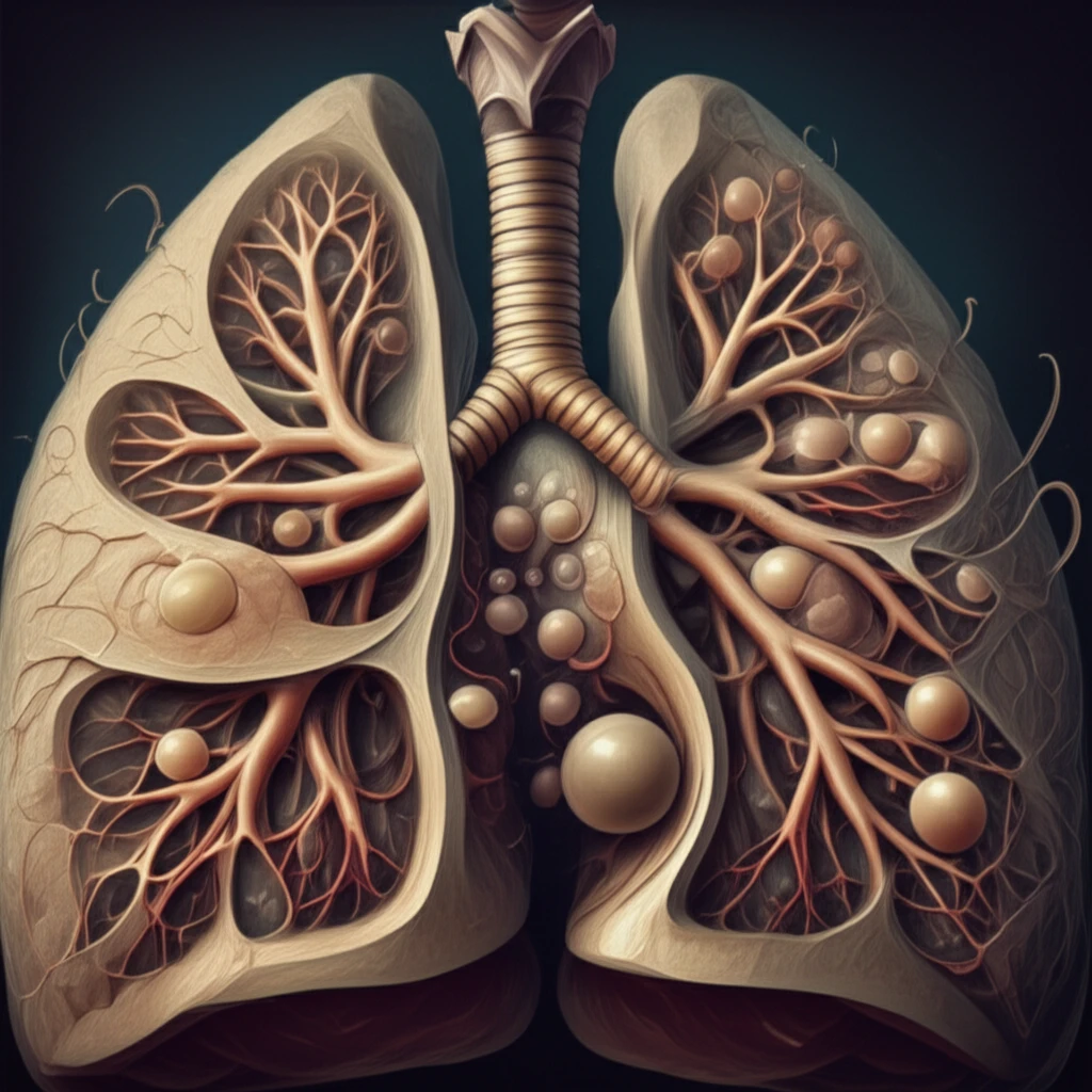
Have you ever heard of a parasitic infection that can take root in your lungs? Pulmonary hydatid cysts, caused by the Echinococcus tapeworm, are a rare but significant health concern, especially in regions where livestock farming is common. Understanding this condition is crucial for early diagnosis and effective treatment.

Hydatid disease, or echinococcosis, is transmitted to humans through contact with infected animals, primarily sheep and cattle. The tapeworm's larval stage forms cysts in various organs, with the liver and lungs being the most common sites. When these cysts develop in the lungs, they can cause a range of respiratory symptoms.

This article delves into a recent case report of a 13-year-old boy diagnosed with pulmonary hydatid cysts, shedding light on the causes, symptoms, diagnosis, and treatment strategies for this often-misunderstood infection.

What Are the Symptoms of Pulmonary Hydatid Cysts?

Illustration of lungs affected by tapeworm cysts

Pulmonary hydatid cysts can manifest in various ways, depending on their size and location in the lungs. Some individuals may experience no symptoms at all in the early stages, while others may develop:

It's important to note that these symptoms can also be indicative of other respiratory conditions, leading to potential misdiagnosis. In the case of the 13-year-old boy, he initially presented with shortness of breath and a non-productive cough, which were initially suspected to be caused by tuberculosis.

  • Chronic cough (either dry or productive)
  • Shortness of breath (dyspnea)
  • Chest pain (pleuritic)
  • Coughing up blood (hemoptysis)
  • In some instances, complications such as bronchial compression or cyst rupture can occur, leading to more severe symptoms. If a cyst ruptures, it can cause allergic reactions or even anaphylaxis.
The young boy also had contact with horse and some cervical lymph node enlargement, but a chest X-ray came back normal, while CT Scans showed two cysts in his left lung.

Early Detection and Treatment Are Key

Pulmonary hydatid cysts, while rare, can pose significant health risks if left untreated. Early detection through imaging techniques like CT scans, combined with accurate diagnosis, is crucial for effective management.

Surgical intervention, involving cyst resection, remains the primary treatment approach. In conjunction with surgery, medications like albendazole are used to prevent recurrence and spread of the infection.

If you experience persistent respiratory symptoms, especially if you have a history of contact with livestock or have lived in endemic regions, consult a healthcare professional for prompt evaluation. Raising awareness and educating communities about the risks of hydatid disease are essential for prevention and control.

About this Article -

This article was crafted using a human-AI hybrid and collaborative approach. AI assisted our team with initial drafting, research insights, identifying key questions, and image generation. Our human editors guided topic selection, defined the angle, structured the content, ensured factual accuracy and relevance, refined the tone, and conducted thorough editing to deliver helpful, high-quality information.See our About page for more information.

This article is based on research published under:

DOI-LINK: 10.4172/2165-7920.1000832, Alternate LINK

Title: Pulmonary Hydatid Cyst In A 13 Years Old Child: A Case Report

Subject: General Medicine

Journal: Journal of Clinical Case Reports

Publisher: OMICS Publishing Group

Authors: Bayan Hafiz, Ashwaq Al Qurani

Published: 2016-01-01

Everything You Need To Know

1

What exactly are Pulmonary hydatid cysts?

Pulmonary hydatid cysts are a rare parasitic infection of the lungs caused by the Echinococcus tapeworm. This infection, known as hydatid disease or echinococcosis, occurs when the larval stage of the tapeworm forms cysts within the lung tissue. These cysts can grow and cause various respiratory symptoms. This condition is particularly relevant in regions where livestock farming, such as sheep and cattle farming, is common, as these animals are the primary hosts for the tapeworm.

2

What are the common symptoms of Pulmonary hydatid cysts?

Symptoms of Pulmonary hydatid cysts can vary depending on the size and location of the cysts within the lungs. In the early stages, some individuals may not experience any symptoms at all. However, as the cysts grow, they can cause a chronic cough, either dry or productive. Other symptoms include shortness of breath (dyspnea), chest pain (pleuritic), and coughing up blood (hemoptysis). It is important to note that these symptoms can mimic those of other respiratory conditions, potentially leading to misdiagnosis. For example, in a reported case, symptoms initially suggested tuberculosis.

3

What causes Pulmonary hydatid cysts?

The Echinococcus tapeworm is the primary cause of Pulmonary hydatid cysts. Humans contract the infection, known as hydatid disease or echinococcosis, through contact with infected animals, especially sheep and cattle. The tapeworm's larval stage develops into cysts that can form in various organs, including the liver and lungs. When these cysts develop in the lungs, they lead to the condition known as Pulmonary hydatid cysts, causing a range of respiratory symptoms depending on the size and location of the cysts.

4

How are Pulmonary hydatid cysts diagnosed?

Diagnosis of Pulmonary hydatid cysts typically involves imaging techniques like CT scans. CT scans are used to visualize the cysts within the lungs, aiding in accurate diagnosis. In the referenced case, a chest X-ray initially came back normal, highlighting the importance of more detailed imaging. Early detection is crucial for effective management, and it helps prevent complications such as bronchial compression or cyst rupture. The case of the 13-year-old boy showed the use of CT scans to identify the cysts in his left lung.

5

Why is early detection and treatment important for Pulmonary hydatid cysts?

Early detection and treatment are critical for managing Pulmonary hydatid cysts effectively. If left untreated, Pulmonary hydatid cysts can lead to complications such as bronchial compression or cyst rupture. A cyst rupture can trigger allergic reactions or even anaphylaxis. Therefore, early diagnosis, often through imaging techniques such as CT scans, allows for timely intervention and reduces the risk of severe outcomes. Effective management strategies are essential to prevent the progression of the disease and minimize health risks.

Newsletter Subscribe

Subscribe to get the latest articles and insights directly in your inbox.