3D printed kidney model over a futuristic operating table

Printing Hope: How 3D Kidney Models Are Changing Healthcare

"From diagnosis to surgical planning, discover how 3D printed kidney models are revolutionizing medical treatment and improving patient outcomes."


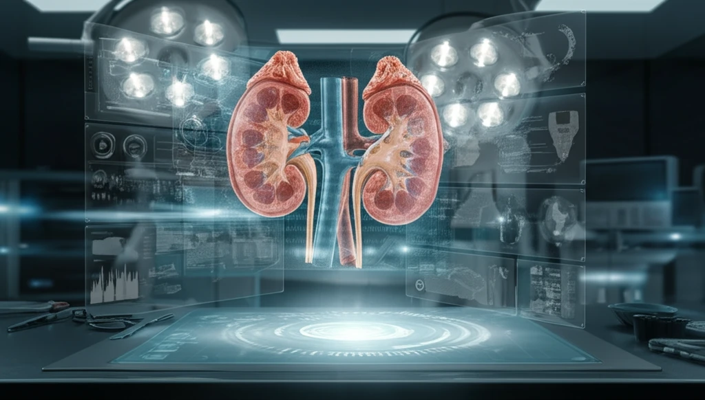
Imagine holding a perfect replica of your kidney in your hands – a tangible model that doctors can use to plan your surgery with unprecedented precision. This isn't science fiction; it's the reality being shaped by 3D printing technology in healthcare. For patients facing kidney-related issues, this innovation offers a beacon of hope, providing clearer insights and potentially improving surgical outcomes.

Traditionally, doctors have relied on CT scans and other imaging techniques to visualize the kidney. However, these methods offer only a two-dimensional view, making it challenging to fully grasp the organ's complex structure, especially when tumors or other abnormalities are present. This is where 3D kidney models come in, bridging the gap between flat images and the three-dimensional reality of the human body.

This article delves into the fascinating world of 3D-printed kidney models, exploring their creation, benefits, and potential to transform healthcare. We'll examine how these models are constructed from CT images, the challenges involved in ensuring accuracy, and the ways in which they are enhancing medical treatment and patient understanding.

Unlocking Precision: How 3D Models of Kidneys Are Made

3D printed kidney model over a futuristic operating table

The journey from a CT scan to a 3D-printed kidney model is a complex yet fascinating process, involving several key steps to ensure accuracy and clinical utility. Here’s how it works:

The process begins with a standard CT scan, which captures detailed cross-sectional images of the patient's kidney. However, raw CT data often suffers from limitations such as lower resolution in the Z-axis (the direction of the scan), which can hinder accurate 3D reconstruction. This is primarily due to radiation risks, where a patient can not be scanned densely. So, CT images are captured at a lower axial resolution.

  • Segmentation: The initial step involves segmenting the kidney region from each CT image. Deep learning methods and contour tracking algorithms are employed to precisely define the kidney's boundaries, distinguishing it from surrounding tissues and organs.
  • 3D Reconstruction: Once the kidney is segmented, the cross-sectional images are stacked to create a preliminary 3D structure. Sophisticated software algorithms then interpolate between the images, compensating for any resolution limitations and generating a smooth, continuous model.
  • Enhancement and Alignment: To further refine the model, interpolation techniques are applied, enhancing details from both cross-sectional and longitudinal views. An alignment scheme registers these views, ensuring accurate representation of the kidney's structure.
  • Vascular Modeling: Beyond the outer structure, 3D kidney models can also incorporate the intricate network of blood vessels within the organ. Vessel tracking and merging schemes reconstruct these inner structures, providing a comprehensive view of the kidney's anatomy.
  • 3D Printing: The final step involves translating the digital model into a physical object using 3D printing technology. Materials such as polymers are carefully selected to mimic the texture and feel of real kidney tissue, allowing surgeons to practice procedures with realistic fidelity.
This meticulous process results in a highly accurate and detailed 3D kidney model that serves as an invaluable tool for surgeons, radiologists, and patients alike. The technology not only reproduces the external structure of the kidney, but also the inner structures like tissues.

The Future of Kidney Care: 3D Printing's Promise

As 3D printing technology continues to advance, its role in kidney care is poised to expand even further. Imagine personalized kidney models tailored to each patient's unique anatomy, enabling surgeons to rehearse complex procedures with unparalleled precision. Picture interactive models that allow patients to visualize their condition and treatment options, empowering them to make informed decisions about their health. These are just a few glimpses of the exciting possibilities that lie ahead. By offering a tangible and detailed representation of the kidney, 3D models are not just improving surgical outcomes; they're also fostering better communication and understanding between doctors and patients, paving the way for a more collaborative and patient-centered approach to healthcare.

About this Article -

This article was crafted using a human-AI hybrid and collaborative approach. AI assisted our team with initial drafting, research insights, identifying key questions, and image generation. Our human editors guided topic selection, defined the angle, structured the content, ensured factual accuracy and relevance, refined the tone, and conducted thorough editing to deliver helpful, high-quality information.See our About page for more information.

This article is based on research published under:

DOI-LINK: 10.1109/ic3.2018.00-23, Alternate LINK

Title: Three-Dimensional Reconstruction And 3D Printing Of Kidney From Computed Tomography

Journal: 2018 1st International Cognitive Cities Conference (IC3)

Publisher: IEEE

Authors: Yu-Zong Huang, Jun-Wei Hsieh, Chian-Hong Lee, Yungchih Chen, Po-Jen Chuang

Published: 2018-08-01

Everything You Need To Know

1

How are 3D kidney models created from CT scans, and what steps are involved in ensuring accuracy?

3D kidney models are created using a multi-step process that begins with a CT scan to capture detailed cross-sectional images of the patient's kidney. The kidney region is then segmented using deep learning methods and contour tracking algorithms to define its boundaries. Next, the cross-sectional images are stacked to create a preliminary 3D structure, and sophisticated software algorithms interpolate between the images to generate a smooth, continuous model. Interpolation techniques are then applied to enhance details from both cross-sectional and longitudinal views, and an alignment scheme registers these views to ensure accurate representation of the kidney's structure. Finally, vessel tracking and merging schemes reconstruct the intricate network of blood vessels within the organ. The digital model is then translated into a physical object using 3D printing technology and materials like polymers to mimic the texture of real kidney tissue.

2

What are the key benefits of using 3D kidney models in medical treatment and surgical planning compared to traditional imaging techniques?

The primary benefit of using 3D kidney models is that they bridge the gap between two-dimensional images, such as CT scans, and the three-dimensional reality of the human body. This allows doctors to grasp the organ's complex structure more fully, especially when tumors or other abnormalities are present. These models provide a tangible and detailed representation of the kidney, improving surgical outcomes and fostering better communication and understanding between doctors and patients. Surgeons can use these models to plan and rehearse complex procedures with unprecedented precision.

3

What are the limitations and challenges in creating accurate 3D kidney models from CT scan data, and how are these addressed?

While the process is detailed, the raw CT data used to create 3D kidney models often suffers from limitations such as lower resolution in the Z-axis, which can hinder accurate 3D reconstruction. This is primarily due to radiation risks, where a patient cannot be scanned densely, so CT images are captured at a lower axial resolution. The segmentation of the kidney region from each CT image requires the use of deep learning methods and contour tracking algorithms to precisely define the kidney's boundaries, distinguishing it from surrounding tissues and organs.

4

How does vascular modeling contribute to the creation of comprehensive 3D kidney models, and why is this level of detail important?

Vascular modeling involves reconstructing the intricate network of blood vessels within the kidney. Vessel tracking and merging schemes are used to reconstruct these inner structures, providing a comprehensive view of the kidney's anatomy. This is important because understanding the vascular structure helps surgeons avoid damaging critical blood vessels during surgery, potentially reducing complications and improving patient outcomes. Incorporating this level of detail allows for a more accurate and realistic surgical planning process.

5

What is the future potential of 3D printing technology in kidney care, and how might it transform patient outcomes and treatment approaches?

The future of 3D printing in kidney care involves personalized kidney models tailored to each patient's unique anatomy, enabling surgeons to rehearse complex procedures with unparalleled precision. Interactive models could allow patients to visualize their condition and treatment options, empowering them to make informed decisions about their health. As 3D printing technology continues to advance, its role in kidney care is poised to expand even further, paving the way for a more collaborative and patient-centered approach to healthcare.

Newsletter Subscribe

Subscribe to get the latest articles and insights directly in your inbox.