Surreal illustration of liver cancer treatment variability

Precision vs. Practicality: Are We Overthinking Liver Cancer Treatment?

"A new study questions the accuracy of current methods for calculating radiation doses in liver cancer treatment, suggesting variability may lead to unexpected outcomes."


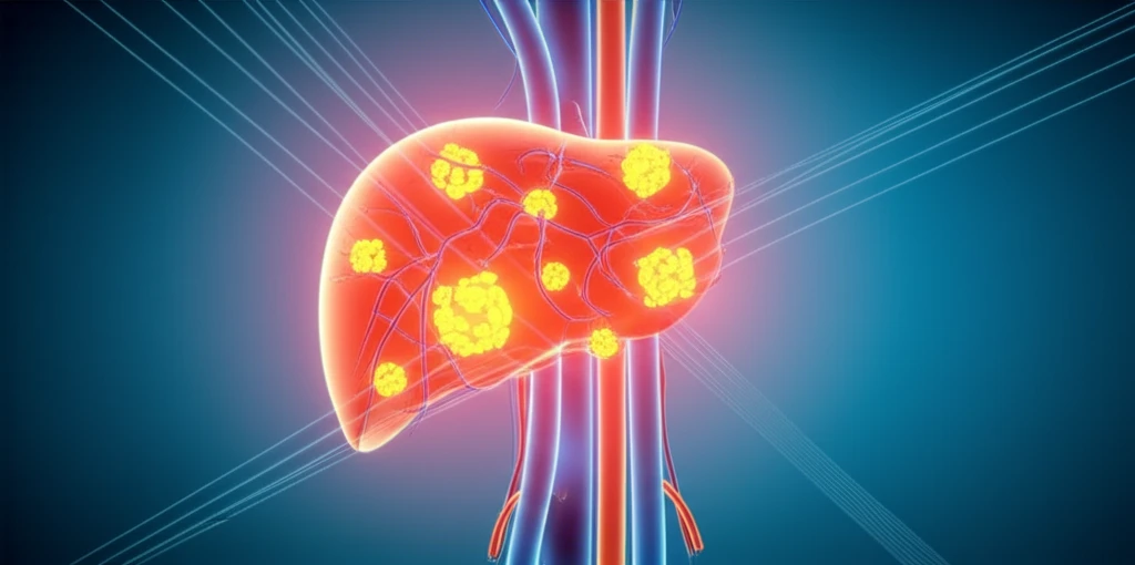
For individuals facing liver cancer, Yttrium-90 radioembolization (Y90 RE) offers a beacon of hope, however, ensuring that the treatment is delivered with utmost precision is critical. The method involves delivering tiny radioactive beads directly to the liver tumors. But how do doctors calculate the correct radiation dose? The answer is more complex than one might think, with potential for significant variability.

Typically, doctors rely on imaging techniques such as CT scans and MRIs to estimate the size of the liver and tumors. These volume measurements are plugged into a formula that determines the appropriate amount of Y90 to administer. A recent study, however, shines a light on the potential for inaccuracies in this process, particularly when compared to cone-beam computed tomography (CBCT), a technique performed during the procedure itself.

The study raises important questions about whether current methods of liver volume calculation are as precise as they need to be, and whether this variability could be leading to unintended consequences in treatment outcomes. It's a reminder that in the world of oncology, even seemingly small discrepancies can have a ripple effect.

Cone-Beam CT vs. Traditional Imaging: What's the Difference?

Surreal illustration of liver cancer treatment variability

The key to understanding the study's findings lies in grasping the difference between CBCT and traditional imaging methods like CT and MRI. While CT and MRI provide detailed anatomical images, they rely on external contrast injected into the body. CBCT, on the other hand, is performed during the actual angiography, allowing doctors to visualize the liver's blood supply in real-time.

This real-time visualization is particularly valuable because it can reveal variations in blood vessel anatomy and areas of extrahepatic perfusion (blood supply to areas outside the liver). These factors can significantly impact how much radiation reaches the tumor and how much is distributed to healthy liver tissue.

Here's a breakdown of the key differences:
  • CBCT: Real-time imaging during angiography, visualizes blood supply, identifies variations in anatomy.
  • CT/MRI: Relies on external contrast, provides detailed anatomical images, may not capture real-time blood flow dynamics.
The study compared liver volume measurements obtained from CBCT images with those from CT and MRI scans. The results revealed that while there wasn't a statistically significant difference in overall volume measurements, there was considerable variability between the two methods. In some cases, the calculated radiation dose differed by more than 50% depending on whether CBCT or traditional imaging was used.

The Path Forward: Balancing Precision and Practicality

This study serves as a reminder that even in advanced medical treatments, there's always room for improvement. While CBCT offers the potential for more precise radiation dose calculations, it also presents practical challenges such as the need for specialized equipment and expertise. The findings highlight the need for ongoing research to refine our methods of liver volume calculation and ensure that patients receive the most effective and safe treatment possible. Further investigation is necessary to see if the variability noted here affects treatment results.

About this Article -

This article was crafted using a human-AI hybrid and collaborative approach. AI assisted our team with initial drafting, research insights, identifying key questions, and image generation. Our human editors guided topic selection, defined the angle, structured the content, ensured factual accuracy and relevance, refined the tone, and conducted thorough editing to deliver helpful, high-quality information.See our About page for more information.

This article is based on research published under:

DOI-LINK: 10.1007/s00270-018-2030-0, Alternate LINK

Title: Comparison Of Cone-Beam Tomography And Cross-Sectional Imaging For Volumetric And Dosimetric Calculations In Resin Yttrium-90 Radioembolization

Subject: Cardiology and Cardiovascular Medicine

Journal: CardioVascular and Interventional Radiology

Publisher: Springer Science and Business Media LLC

Authors: Marco Ertreo, Hailey Choi, David Field, Jonathan W. Lischalk, Emil Cohen, George E. Lynskey, Theresa Caridi, Donna Buckley, Keith Unger, Alexander Y. Kim

Published: 2018-07-13

Everything You Need To Know

1

What is Yttrium-90 radioembolization (Y90 RE), and why is it important in the context of liver cancer treatment?

Yttrium-90 radioembolization (Y90 RE) is a treatment for liver cancer that delivers tiny radioactive beads directly to liver tumors. It is a crucial method for individuals facing liver cancer, offering a beacon of hope. The main implication is delivering precise radiation dose, which is critical for the success of the treatment, making it a vital procedure in the field of oncology.

2

How are radiation doses calculated in Yttrium-90 radioembolization (Y90 RE), and why is precision important in this process?

The primary methods used for calculating radiation doses for Y90 RE involve estimating liver and tumor sizes using imaging techniques such as CT scans and MRIs. These volume measurements are then input into a formula to determine the amount of Y90 to administer. The study highlights the importance of accurate measurements for calculating the appropriate dose of Y90 for effective treatment, and any inaccuracies could lead to unintended consequences in treatment outcomes.

3

What is the difference between Cone-Beam CT (CBCT) and traditional imaging methods like CT and MRI, and what are the implications of each in liver cancer treatment?

CBCT is a real-time imaging technique performed during angiography, allowing doctors to visualize the liver's blood supply in real-time, unlike CT and MRI, which rely on external contrast. CBCT's real-time visualization reveals variations in blood vessel anatomy and areas of extrahepatic perfusion. The key implication here is its ability to refine the method of liver volume calculation to ensure patients receive the most effective and safe treatment possible, which is not fully achievable with CT and MRI.

4

What did the study reveal about the variability in liver volume measurements between CBCT and traditional imaging?

The study found variability in liver volume measurements between CBCT and traditional imaging methods, such as CT and MRI, despite no statistically significant difference in overall volume measurements. The variability could lead to calculated radiation doses differing by over 50%, depending on the imaging method used. This suggests that current methods might not be as precise as needed, which underscores the importance of refining these methods for more accurate and effective treatment.

5

What are the next steps in refining liver cancer treatment methods, based on the study's findings?

The path forward involves balancing the potential for more precise radiation dose calculations using methods like CBCT with the practical challenges, such as the need for specialized equipment and expertise. Ongoing research is necessary to refine methods of liver volume calculation and improve patient outcomes. Further investigation is necessary to see if the variability noted here affects treatment results.

Newsletter Subscribe

Subscribe to get the latest articles and insights directly in your inbox.