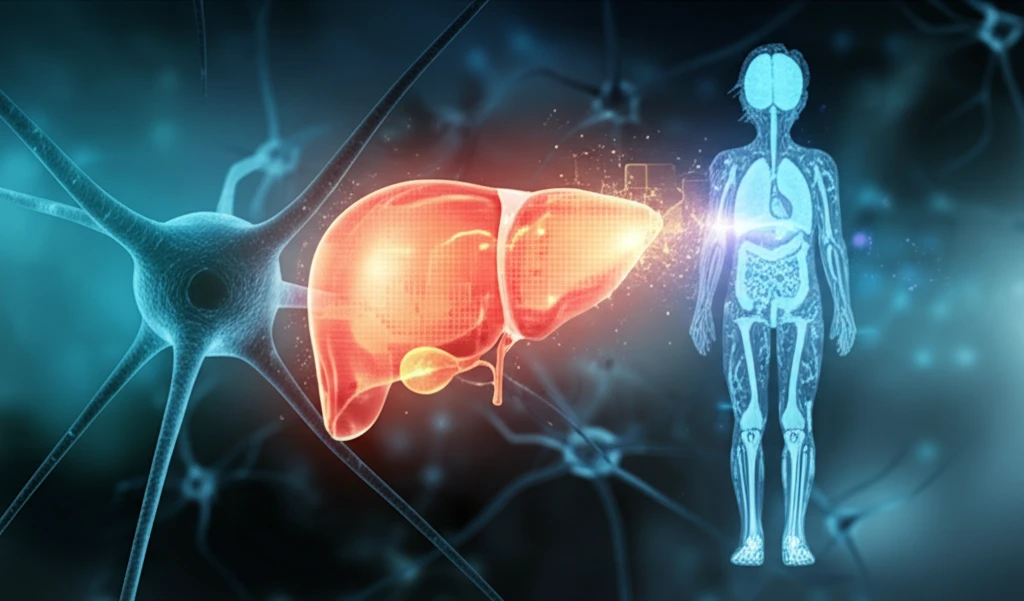
AI-enhanced liver segmentation for children

Precision in Paediatrics: The Quest for Sharper Liver Scans

"New AI Advances Promise Safer, More Accurate Medical Imaging for Children"


The delicate nature of paediatric healthcare requires a constant balance between diagnostic accuracy and patient safety. When it comes to medical imaging, children are particularly vulnerable to the harmful effects of radiation. This is why combined PET-CT scanners, which offer lower radiation doses, are increasingly used. However, the resulting CT images often suffer from low contrast, making it challenging for doctors to accurately segment and analyze the liver.

Imagine trying to find a single puzzle piece in a box where all the pieces are the same color. That's the challenge doctors face when trying to segment a child's liver in a low-contrast CT scan. Accurate segmentation is crucial for diagnosis, treatment planning, and monitoring disease progression. Manual segmentation is time-consuming and prone to human error. This has spurred researchers to develop automated methods that can tackle the challenge effectively.

The field of liver segmentation has seen remarkable progress, with techniques ranging from probabilistic atlases to statistical shape models. Yet, the unique challenges posed by low-contrast paediatric images demand innovative solutions. A new study introduces an adaptive kernel-based Statistical Region Merging (SRM) algorithm, offering a promising step forward in the quest for precise and safe liver segmentation in children.

Adaptive Kernel-Based SRM: A Closer Look

AI-enhanced liver segmentation for children

The core of this innovation lies in the adaptive kernel-based Statistical Region Merging (SRM) algorithm. It’s a mouthful, but the concept is ingenious. The algorithm is designed to address the issue of low contrast by merging pixels into statistically homogenous regions. The algorithm then leverages an adaptive kernel that sharpens image clarity to allow the automated systems to detect differences more readily. This enhancement is critical for accurately defining the liver's boundaries. The kernel adapts to the characteristics of each image, effectively smoothing out noise and enhancing relevant features.

Think of it like noise-cancelling headphones for medical images. The adaptive kernel identifies and filters out the 'noise' (low contrast and artifacts) that obscures the liver's details, allowing the algorithm to focus on the essential structures. The kernel function is determined by automatically analyzing the noise distribution in each CT image, which allows it to be fine-tuned to the unique characteristics of the scan.

The key benefits of the adaptive kernel-based SRM algorithm include:
  • Improved Accuracy: Achieves a higher Dice index compared to traditional SRM methods.
  • Enhanced Robustness: Successfully segments all CT images, even those with significant low contrast.
  • Reduced Manual Intervention: Automates the segmentation process, saving time and resources.
  • Personalized Adaptation: Adapts to the unique noise characteristics of each image.
In essence, this adaptive method represents a significant step toward more reliable and efficient liver segmentation in pediatric patients. By addressing the limitations of low-contrast imaging, it enhances diagnostic precision and minimizes the need for invasive procedures. This reduces risks involved with exploration and helps give doctors more information for treatment.

The Future of Paediatric Liver Imaging

While these results are promising, the researchers acknowledge that further validation with a larger dataset is essential. This highlights the ongoing need for collaborative efforts to refine and expand the applications of AI in paediatric medical imaging. The journey toward safer, more accurate diagnostics for children is a continuous one, and innovations like the adaptive kernel-based SRM algorithm pave the way for a brighter future.

About this Article -

This article was crafted using a human-AI hybrid and collaborative approach. AI assisted our team with initial drafting, research insights, identifying key questions, and image generation. Our human editors guided topic selection, defined the angle, structured the content, ensured factual accuracy and relevance, refined the tone, and conducted thorough editing to deliver helpful, high-quality information.See our About page for more information.

This article is based on research published under:

DOI-LINK: 10.1007/978-3-030-00807-9_17, Alternate LINK

Title: Paediatric Liver Segmentation For Low-Contrast Ct Images

Journal: Data Driven Treatment Response Assessment and Preterm, Perinatal, and Paediatric Image Analysis

Publisher: Springer International Publishing

Authors: Mariusz Bajger, Gobert Lee, Martin Caon

Published: 2018-01-01

Everything You Need To Know

1

Why are low-contrast CT images a problem in pediatric liver segmentation?

In pediatric liver segmentation, low-contrast CT images pose a significant problem because they make it difficult for doctors to accurately define the boundaries of the liver. This lack of clear definition complicates diagnosis, treatment planning, and the monitoring of disease progression. The low contrast arises due to the use of lower radiation doses in combined PET-CT scanners, which are favored to minimize radiation exposure in children.

2

How does the adaptive kernel-based Statistical Region Merging (SRM) algorithm improve liver segmentation?

The adaptive kernel-based Statistical Region Merging (SRM) algorithm enhances liver segmentation by merging pixels into statistically homogenous regions, addressing the issue of low contrast in CT images. It uses an adaptive kernel to sharpen image clarity, enabling automated systems to better detect differences and define the liver's boundaries. The kernel effectively smooths out noise and enhances relevant features, adapting to the unique characteristics of each image by analyzing the noise distribution.

3

What are the key benefits of using the adaptive kernel-based Statistical Region Merging (SRM) algorithm for pediatric liver scans?

The adaptive kernel-based Statistical Region Merging (SRM) algorithm improves accuracy in liver segmentation, achieving a higher Dice index compared to traditional SRM methods. It also enhances robustness by successfully segmenting all CT images, even those with significant low contrast, while reducing manual intervention through automation, saving time and resources. Additionally, its personalized adaptation tailors to the unique noise characteristics of each image, ensuring optimal performance across diverse scan qualities.

4

What are the next steps in developing the adaptive kernel-based Statistical Region Merging (SRM) algorithm for pediatric liver imaging?

While the adaptive kernel-based Statistical Region Merging (SRM) algorithm shows promise in improving liver segmentation in low-contrast CT images, the next steps involve validation with a larger dataset. Further research is needed to explore the full potential and refine the applications of AI in paediatric medical imaging to ensure safer and more accurate diagnostics for children. Continuous efforts and collaboration are essential to advancing this technology.

5

What are probabilistic atlases and statistical shape models, and how do they relate to the adaptive kernel-based Statistical Region Merging (SRM) algorithm?

Probabilistic atlases and statistical shape models represent earlier techniques used in liver segmentation. They are approaches that use prior knowledge about the typical shape and appearance of the liver to guide the segmentation process. However, these methods often struggle with the unique challenges posed by low-contrast paediatric images, which require more innovative solutions like the adaptive kernel-based Statistical Region Merging (SRM) algorithm to effectively address noise and enhance image clarity.

Newsletter Subscribe

Subscribe to get the latest articles and insights directly in your inbox.