Precision in Cancer Treatment: 3D Dose Measurement Breakthrough
"New method uses Fricke gel dosimeters and optical computed tomography to enhance HDR brachytherapy, improving accuracy and reducing side effects."
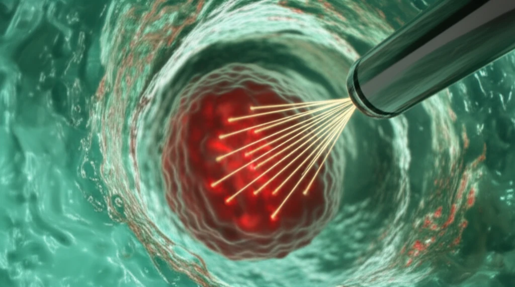
High-dose-rate (HDR) brachytherapy is a precise cancer treatment that places radioactive sources directly inside or next to tumors. This method allows doctors to deliver high doses of radiation to cancer cells while minimizing damage to surrounding healthy tissue. The key to its effectiveness lies in the accuracy of the dose delivery, which ensures that the tumor receives the intended radiation while sparing critical organs.
Measuring radiation dose distribution accurately, especially around HDR brachytherapy sources, is challenging. Traditional dosimetry systems often lack the necessary spatial resolution and sensitivity to capture the sharp dose gradients near the radiation source. This limitation has spurred the development of advanced techniques to visualize and verify the dose distribution in three dimensions.
A promising solution involves Fricke gel dosimeters combined with optical computed tomography (CT). Fricke gels react to radiation by changing color, and optical CT scans can then create detailed 3D images of these color changes, revealing the dose distribution. This method offers high spatial resolution and is nearly water-equivalent, meaning it doesn't significantly alter the radiation field. Recent research has refined this technique to overcome challenges, paving the way for more accurate and reliable HDR brachytherapy treatments.
How Fricke Gel Dosimeters and Optical CT Scanning Work Together
The recent study published in 'Australasian Physical & Engineering Sciences in Medicine' details a new method for enhancing the precision of HDR brachytherapy using Fricke gel dosimeters and optical CT. The researchers aimed to create an accurate, high-resolution technique for measuring 3D dose distributions, addressing limitations of previous methods.
- Phantom Creation: A custom phantom was 3D-printed to hold brachytherapy needles in a precise arrangement.
- Gel Preparation: Fricke Xylenol gel was carefully prepared and poured into the phantom, encasing the needles.
- Treatment Planning: The HDR brachytherapy treatment was planned using specialized software to deliver a specific dose distribution.
- Irradiation: The gel phantom was irradiated with the planned treatment, causing chemical changes in the gel proportional to the radiation dose.
- Optical CT Scanning: After irradiation, the needles were removed and replaced with a refractive index-matched fluid to minimize artifacts during scanning. The gel was then scanned using optical CT to create a 3D image of the dose distribution.
- Data Analysis: The CT images were processed to subtract background noise and convert optical density changes into dose values. These measured dose values were then compared to the planned dose distribution.
The Future of HDR Brachytherapy
The phantom construction and optical CT scanning method offer the potential for routine quality assurance measurements of complex HDR brachytherapy treatment deliveries. By adapting this technique, clinicians can ensure that radiation is delivered precisely as planned, optimizing treatment outcomes and minimizing side effects. This innovation opens new avenues for personalized cancer therapy, where treatment plans are verified with unprecedented accuracy.